Lumbar MRIs are likely the most common MRI people would report but to see acute denervation changes from disc herniation is uncommon. So what does denervation look like in the lumbar spine?
In the acute to sub-acute stage there is an " oedema" type pattern to the signal with increased T2 signal, iso-intense T1 signal and enhancement of the affected muscle/s post contrast. Muscle atrophy and fatty infiltration is progressive and as this case shows, can be quite significant even within 2 weeks.
In the chronic stage the "oedema" type signal resolves and there is muscle atrophy and fatty infiltration.
This man presented with sciatica after coughing and the scan was done about two weeks after onset of symptoms.
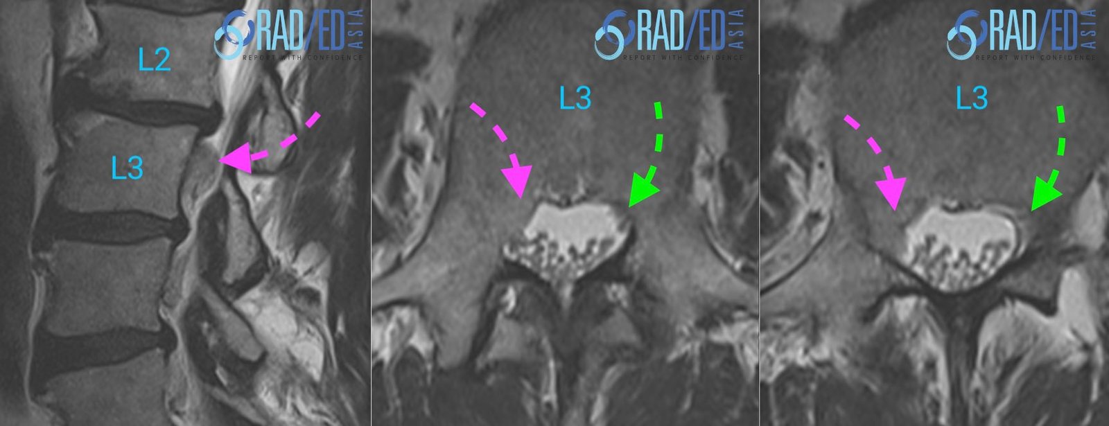
Image above: Right paracentral disc extrusion (Pink arrow) at L2/3 compressing the traversing right L3 nerve root (Pink arrow axials.).Normal left L3 traversing nerve root Green arrow.
DENERVATION CHANGES:

Image above: Compare both sides to appreciate the loss of volume, increased T2 signal and fatty change on the right posterior paraspinal muscles. Easy to miss if we concentrate just on the canal.
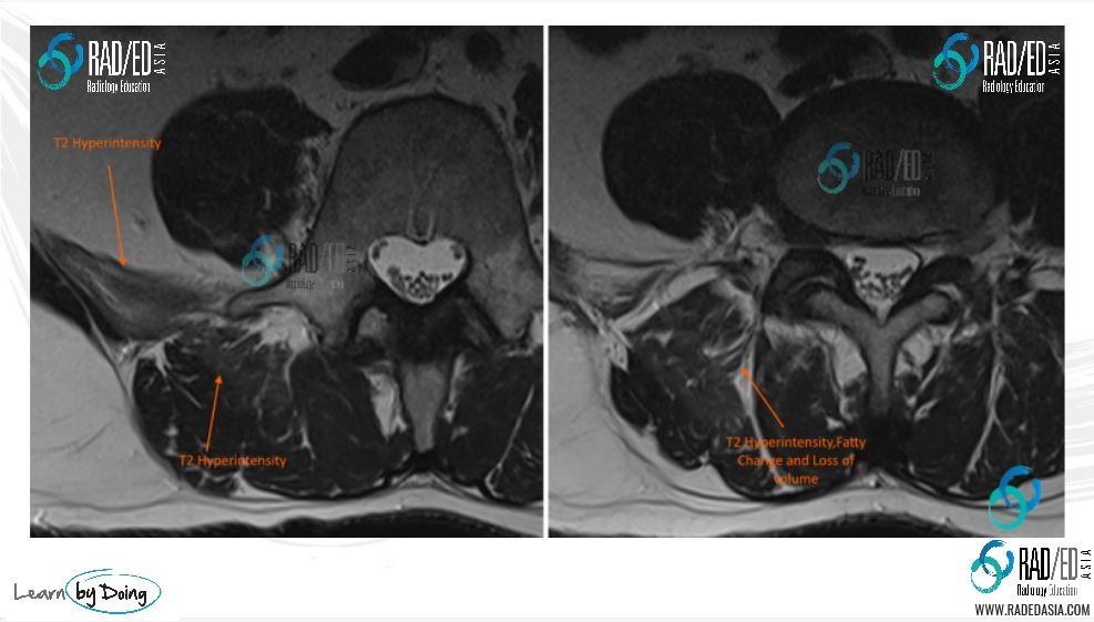
Image above: T2 scans: Compare both sides to appreciate the loss of volume, particularly of quadratus lumborum (anterior arrow) increased T2 signal and fatty change on the right posterior paraspinal muscles.
It's not common to give contrast for denervation but if you do, expect to see enhancement in the acute and subacute phase.

Image Above: Post contrast scans demonstrate enhancement of denervated muscles paraspinal and right quadratus lumborum. .
- So in this patient 2 weeks after the acute disc prolapse we see muscle oedema, loss of volume with fatty infiltration and enhancement of denervated muscles.
- The T2 hyper-intensity is easy to pass by if you are not looking carefully and would be more obvious if a T2 fat sat or STIR scan was performed. On the post contrast scans though the signal abnormality is much easier to appreciate.
- As lumbar imaging is mostly without contrast and without a T2 FS or STIR sequence its quite easy to not notice denervation changes if we concentrate just on the canal when reporting.


#radedasia #mri #mskmri #radiology