Dr Ravi Padmanabhan

neuropathic joint-syrinx syringomyelia spine mri online radiology courses

NEUROPATHIC JOINT ELBOW SYRINX SYRINGOMYELIA SPINE MRI

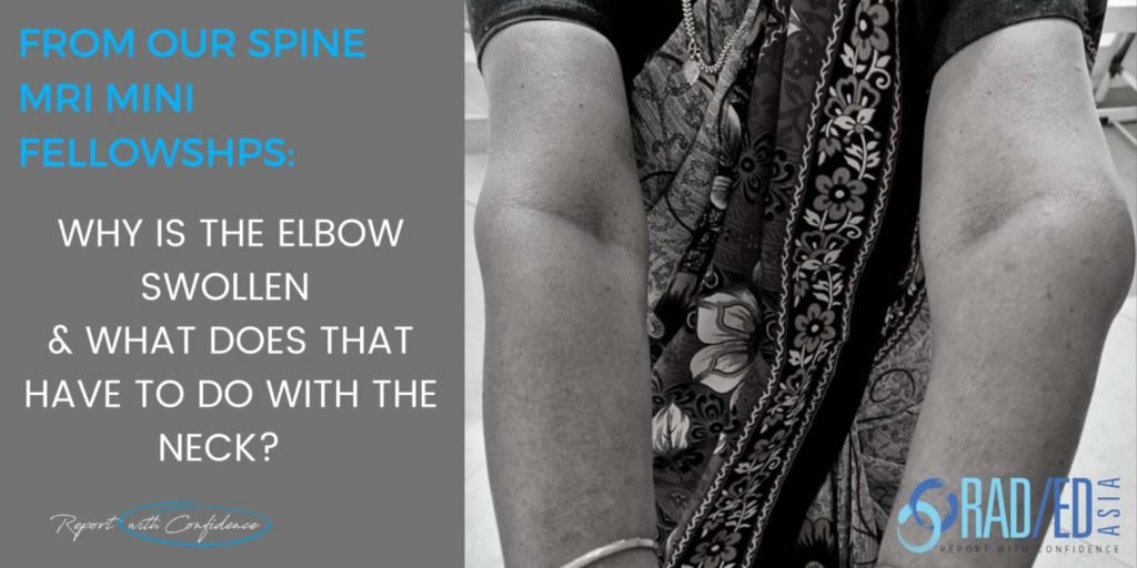
NEUROPATHIC JOINT ELBOW SYRINGOMYELIA SPINE MRI A combined Clinico Radiological case from Dr Joe Thomas (Rheumatologist) Neuropathic Elbow Joint due to a Syrinx. A case from Dr Joe Thomas a senior consultant Rheumatologist who is also joining us in presenting the Spine Arthropathy and Spondyloarthropathy course. CLINICO RADIOLOGICAL CORRELATION CLINICAL PRESENTATION Right handed 60 yo …

NEUROPATHIC JOINT ELBOW SYRINX SYRINGOMYELIA SPINE MRI Read More »

shoulder knee mri msk radiology kuala lumpur workshop conference course musculoskeletal imaging

MSK RADIOLOGY CONFERENCE COURSE MRI KNEE SHOULDER MUSCULOSKELETAL KUALA LUMPUR MALAYSIA

Home MSK MRI RADIOLOGY CONFERENCE COURSE MUSCULOSKELETAL MALAYSIA KUALA LUMPUR: MSK MRI RADIOLOGY KNEE & SHOULDER MINI FELLOWSHIP: 16/17 SEPTEMBER 2023 This Mini Fellowship is now over.To see our latest Onsite Guided Mini Fellowships please click HERE FROM THE MINI-FELLOWSHIP @ KL Our 2-Day Onsite Knee and Shoulder MRI Mini Fellowship and Workstation Workshop will …

MSK RADIOLOGY CONFERENCE COURSE MRI KNEE SHOULDER MUSCULOSKELETAL KUALA LUMPUR MALAYSIA Read More »

mri cystic arachnoiditis cord compression oedema radiology education asia

MRI CORD OEDEMA FROM CYSTIC ARACHNOIDITIS

MRI CORD OEDEMA FROM CYSTIC ARACHNOIDITIS CORD OEDEMA FROM CYSTIC ARACHNOIDITIS Spinal arachnoiditis can have various appearances. One of the more severe forms, cystic arachnoiditis, can result in cord signal abnormality which can be very extensive. This post looks at why cord oedema develops in cystic arachnoiditis and its MRI appearance. PATHOLOGY Normally there is …

MRI CORD OEDEMA FROM CYSTIC ARACHNOIDITIS Read More »

learn-elbow-imaging-mri-radiology-online-course-radedasia

ELBOW MRI RADIOLOGY MSK ONLINE COURSE GUIDED LEARNING MAY 2024

Home MRI ELBOW ONLINE MINI FELLOWSHIP (GUIDED) COURSE BEGINS 4th MAY 2024 Our new MRI ELBOW Online radiology Course will cover the common abnormalities we see in reporting, focusing on how we assess a scan at work … What to Look for, Where to Look and How to Best Report it. The focus of this Online MRI Elbow course, like all …

ELBOW MRI RADIOLOGY MSK ONLINE COURSE GUIDED LEARNING MAY 2024 Read More »

elbow mri brachialis tendon tears tendinosis radiology education asia

MRI ELBOW: MRI BRACHIALIS TENDON TEARS, TENDONOSIS AND NORMAL

MRI BRACHIALIS TENDON BRACHIALIS TENDON MRI: Elbow MRI Brachialis tendon Tendinosis, Tears and Normal   The Brachialis tendon is less commonly injured than the biceps.  It inserts onto the anterior ulnar on the ulnar tuberosity and to a lesser extent on the coronoid process but the tendon is very short compared to the biceps tendon. …

MRI ELBOW: MRI BRACHIALIS TENDON TEARS, TENDONOSIS AND NORMAL Read More »

itb iliotibial band friction syndrome bursa radiology mri knee

ITB ILIOTIBIAL BAND FRICTION SYNDROME MRI: IS IT A BURSA OR A RECESS?

ITB ILIOTIBIAL BAND FRICTION SYNDROME MRI TWO POSSIBILLITIES With fluid deep to the ITB we have 2 possibilities.  It’s a recess of the suprapatellar bursa or, It’s an ITB bursa (an adventitial bursa which develops due to friction). HOW DOES THE ANATOMY HELP TO DIFFERENTIATE? ITB BURSA will NOT communicate with the suprapatellar recess or …

ITB ILIOTIBIAL BAND FRICTION SYNDROME MRI: IS IT A BURSA OR A RECESS? Read More »

This site is intended for Medical Professionals only. Use of this site is governed by our Terms of Service and Privacy Statement which can be found by clicking on the links. Please accept before proceeding to the website.
error: Right Click not available.