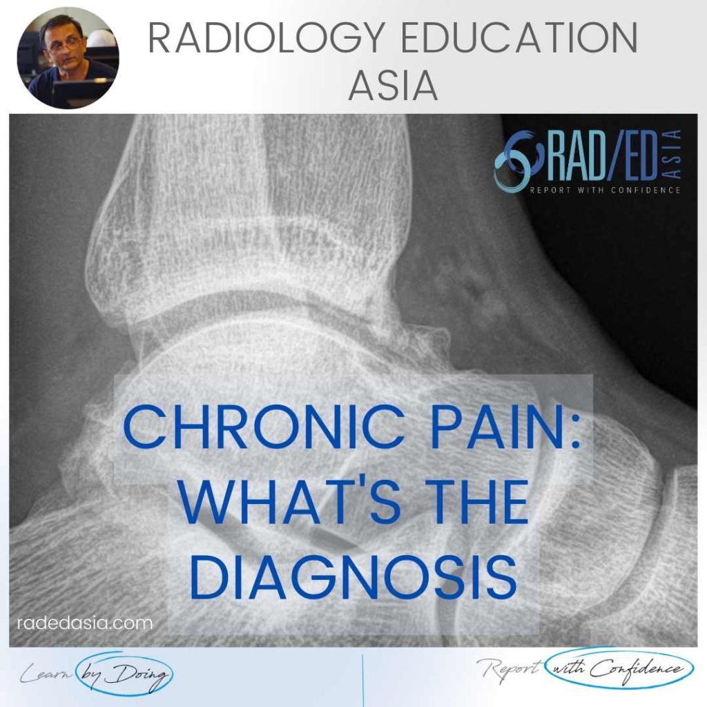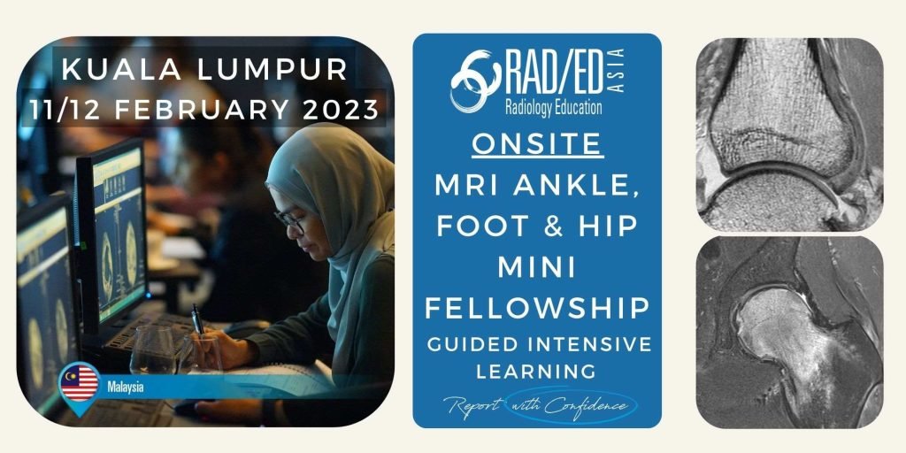
MSK RADIOLOGY CONFERENCE COURSE MUSCULOSKELETAL MRI ANKLE FOOT & HIP MALAYSIA KUALA LUMPUR
Home MSK RADIOLOGY CONFERENCE COURSE MUSCULOSKELETAL MRI MALAYSIA KUALA LUMPUR: MSK MRI RADIOLOGY ANKLE, FOOT & HIP MINI FELLOWSHIP: 11/12 FEBRUARY 2023 This Mini Fellowship is now over.To see our latest Onsite Guided Mini Fellowships please click HERE WHAT HAPPENED AT THE MINI-FELLOWSHIP @ KL CLICK ON THE IMAGE BELOW TO FIND OUT Our 2-Day …