radiology conference mri fellowship course

MRA SHOULDER ARTHROGRAMS: HOW TO DO IT

Shoulder arthrograms for MRI are very useful to better see labral or SLAP tears especially if you have a low field strength magnet or you are starting and don’t have enough experience. They are easy to do and add a lot to the accuracy for diagnosing labral tears. Here is how to do them. The post is in response to a question from one of the radiologists at the recent workshop on how to do them.

1. Patient prone arm extended palm up and put a sand bag on the palm to hold it externally rotated.

shoulder
2. Mark position at supero medial corner of humeral head around level of coracoid. Mark over the bone and not at the joint space.

shoulder arthrogram mri mra radiology education asia

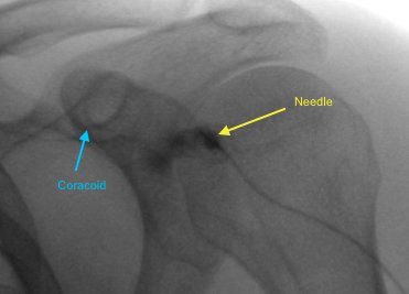
Why at level of coracoid tip and not lower at inferior half of humeral head?
 
shoulder arthrogram mri radiology education asia
 
shoulder arthrogram mri subscapularis radiology education asia
 Images Above: Low entry site will go through subscapularis tendon which can feel very firm and you think you have hit bone. But when you inject its extra articular still. Injecting at the level of the coracoid process avoids this.

3. Sterile technique 22 gauge needle (make sure you have a sharp tip needle like a Terumo needle. The standard spinal needles don’t cut well) push it vertically down over the marked spot until you feel bone. Sometimes subscap tendon can feel very firm so push until it won’t go further.
 
4. 3 ml syringe with iodinated contrast connected to a minimal volume tube. Connect to needle and inject under fluoro screening. Contrast should flow away either into sub-coracoid space (like image above) or into joint and inferiorly. There shouldn't be any resistance to injecting.
 
5. Once you have confirmed you are in joint, inject diluted gadolinium or saline (up to 10 mls).

shoulder arthrogram mri gadolinium radiology education asia

  • If using Gadolinium:

 
shoulder arthrogram mri gadolinium mra radiology education asia

     
  • If using Saline: 
    3 planes PD and PDFS
Posterior approach arm adducted and ipsilateral hand on contralateral shoulder. Insert needle to posterior joint and inject 1-2mls of saline or local anesthetic to make sure you are in joint (see it flowing in joint and no resistance to injection). Once you have confirmed position, inject gadolinium/ saline as above.

Learn more about SHOULDER Imaging in our ONLINE or ONSITE
Guided MRI SHOULDER Mini-Fellowship.


More by clicking on the images below.

#radiology #radedasia #radiologia #msk #mskmri #shouldemri #mra #arthrogram #rheumatology #arthritis #rheumatologist #sportsphysician #orthopaedic #chiropractic #sportsmedicine #orthopaedic

#radedasia #mri #mskmri #radiology

This site is intended for Medical Professionals only. Use of this site is governed by our Terms of Service and Privacy Statement which can be found by clicking on the links. Please accept before proceeding to the website.
error: Right Click not available.