
Identifying the gleno humeral ligaments on MRI can be challenging as they are small and on lower field strength scanners there is inadequate resolution to see them properly.
However if we understand the anatomy of the ligaments it becomes easier to know where to look even if we cant directly identify the ligament. This is the 2nd in the series (First post on the Superior Gleno Humeral Ligament click on this link SGHL
This post is an adaptation of a talk given at Radiology Asia in Singapore titled Gleno Humeral Ligaments made Easy…er.![]()
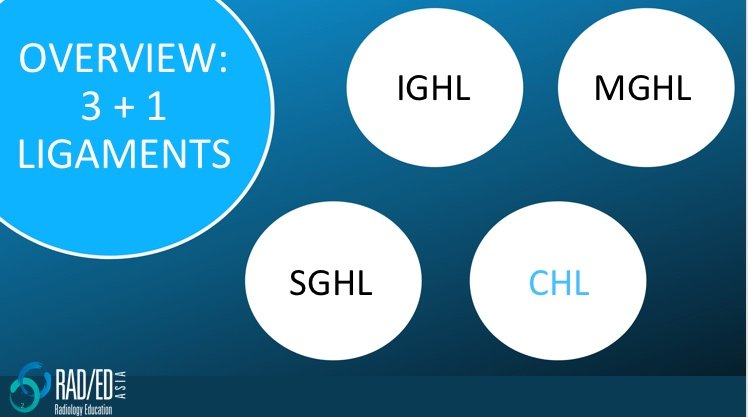
There are 3 main Glenohumeral ligaments.
1. SGHL: Superior GlenoHumeral Ligament (Click here for more on the SGHL )
2. MGHL: Middle GlenoHumeral Ligament
3. IGHL: Inferior GlenoHumeral Ligament
4. The Coraco Humeral Ligament (CHL) is extra capsular and is not part of the Glenohumeral ligaments but we will also look at as it helps to identify the SGHL and it also fuses with the joint capsule at the rotator interval. (Click here for more on the CHL)
![]()
The ligaments arise in an orderly fashion and using analogies often helps to understand anatomy. On sagittal scans the ligaments have the appearance of tentacles of an octopus radiating from the biceps insertion on the supra glenoid tubercle.
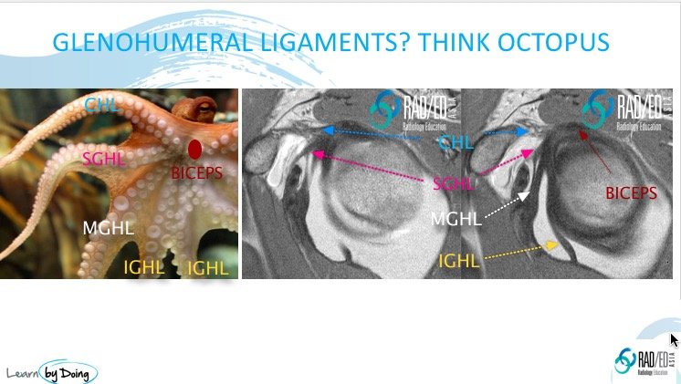
After having identified the CHL (Blue arrow) and SGHL (Pink arrow), the next structure that you see immediately below it on sagittal scans is the MGHL (White arrow).![]()

- The MGHL arises most commonly from the antero superior labrum or neck of glenoid.
- It can be bifid.
- Its best seen on axial scans.
- Look for a linear structure (Blue arrow) running anterior to the labrum (Pink arrow) which eventually inserts into the anterior capsule (Yellow arrows) posterior to subscapularis.
![]()
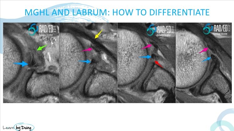
Image above: Two black, linear structures. Which is the labrum and which is the MGHL?
- The key to differentiating the MGHL from a torn labrum is to follow the two structures from superior to inferior on the axial scans.
- The MGHL (Pink arrow) will be more anterior but most importantly can be followed inferiorly to its attachment with the anterior capsule posterior to subscapularis (Yellow arrow).
- The labrum (Blue arrow) whilst displaced, is more posterior and as you scroll inferiorly, does not attach to the anterior capsule.
- Green arrow (Torn proximal MGHL which is ill defined and hyper-intense).
- Red Arrow (tear through the base of the anterior labrum).
![]()


#radedasia #mri #mskmri #radiology