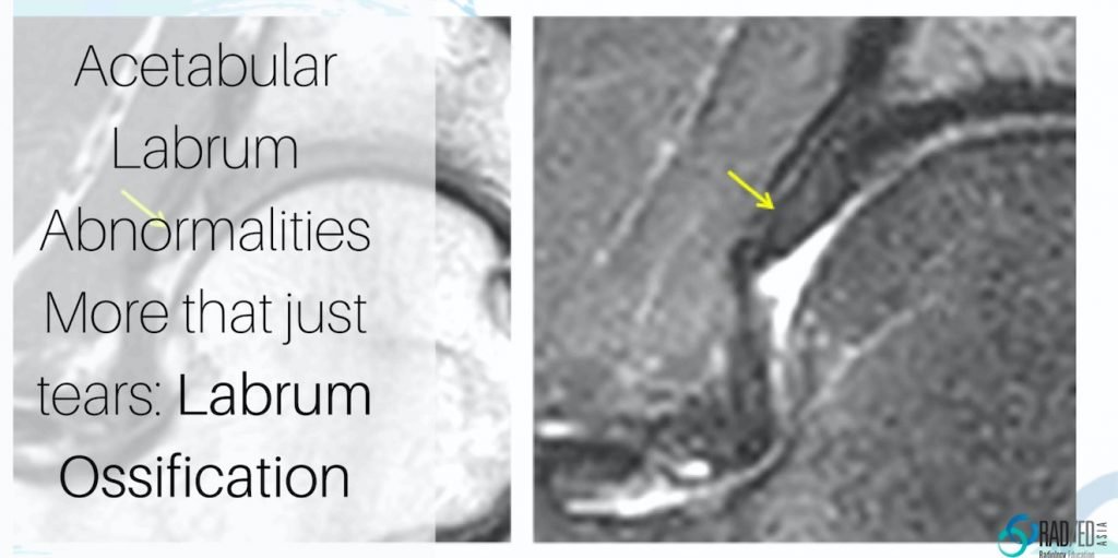
Dr Ravi Padmanabhan

axillary-nerve-denervation-quadrilateral-space-syndroma-dislocation-radiology-education-asia-radedasia

MRI SHOULDER DENERVATION: QUADRILATERAL SPACE SYNDROME AND DISLOCATION

Denervation around the Shoulder: Quadrilateral Space Syndrome and Dislocation.   This post looks at denervation of the Axillary Nerve in  Quadrilateral Space Syndrome and Axillary nerve damage post shoulder dislocation both of which are less common causes of denervation around the shoulder. They have a particular distribution so recognizing the muscles involved gives you the …

MRI SHOULDER DENERVATION: QUADRILATERAL SPACE SYNDROME AND DISLOCATION Read More »

learn mri radiology hip labrum mucoid degen radedasia

HIP MRI LABRUM MUCOID DEGENERATION (VIDEO)

HIP LABRUM MUCOID DEGENERATION MUCOID DEGENERATION MRI APPEARANCE This is the MRI appearance of mucoid degeneration of the hip labrum. On MRI look for a number of findings Enlargement of the labrum Hyper-intensity of the labrum There may or may not be intra labral cysts of varying size. FINDINGS OF MUCOID DEGENERATION MRI Coronal PDFS …

HIP MRI LABRUM MUCOID DEGENERATION (VIDEO) Read More »

mri ankle foot ligament short plantar

MRI PLANTAR LIGAMENT SHORT PLANTAR LIGAMENT ANKLE FOOT AND TOE (VIDEO)

MRI PLANTAR LIGAMENT SHORT PLANTAR LIGAMENT WHAT'S THE STRUCTURE The Short Plantar Ligament. WHAT ARE THE FINDINGS ANATOMY OF THE SHORT PLANTAR LIGAMENT. 1. Extends from the calcaneus (Green star) to the cuboid (Yellow star). 2. It is distal and separate to long plantar ligament. 3. Has a fan shaped appearance. MRI PLANTAR LIGAMENT SHORT …

MRI PLANTAR LIGAMENT SHORT PLANTAR LIGAMENT ANKLE FOOT AND TOE (VIDEO) Read More »

subcortical subchondral fracture knee tibial plateau mri insufficiency

SUBCORTICAL SUBCHONDRAL FRACTURE KNEE MRI TIBIAL PLATEAU (VIDEO)

SUBCORTICAL SUBCHONDRAL FRACTURE KNEE MRI TIBIAL PLATEAU DISCUSSION Subchondral or subcortical fractures of the knee can occur in the femoral condyles or tibial plateau.   The key to the diagnosis is seeing a linear low signal line adjacent to and paralleling the cortex without any cortical defect or break in the acute phase.     There …

SUBCORTICAL SUBCHONDRAL FRACTURE KNEE MRI TIBIAL PLATEAU (VIDEO) Read More »

mri fellowship msk spine musculoskeletal radiology radedasia

HOW WILL YOU LEARN IN THE ONSITE AND ONLINE GUIDED MINI FELLOWSHIPS

HOW WILL YOU LEARN IN THE ONSITE AND ONLINE GUIDED MINI FELLOWSHIPS These are three videos of How you will Learn in both our Onsite and Online Mini Fellowships . The 1st is an Overview of Both. 2nd is How the Online Mini Fellowships are run. 3rd is What are some of the new methods …

HOW WILL YOU LEARN IN THE ONSITE AND ONLINE GUIDED MINI FELLOWSHIPS Read More »

mri ct osteitis condensans ilii spine sij

OSTEITIS CONDENSANS ILII RADIOLOGY CT SACROILIAC JOINT SIJ (VIDEO)

OSTEITIS CONDENSANS ILII RADIOLOGY CT SACROILIAC JOINT SIJ DISCUSSION ON OCI Osteitis Condensans Ilii.   Typical CT sclerotic changes of Osteitis Condensans Ilii. The sclerosis is continuous with the SIJ margin and particularly on the patient’s right it has a triangular appearance.   There is also mild sclerosis in the right sacrum adjacent to the …

OSTEITIS CONDENSANS ILII RADIOLOGY CT SACROILIAC JOINT SIJ (VIDEO) Read More »

iliotibial band mri anatomy syndrome trauma mri knee radedasia

ILIOTIBIAL BAND MRI ANATOMY, SYNDROME, TRAUMA KNEE RADIOLOGY

ILIOTIBIAL BAND MRI ANATOMY, SYNDROME, TRAUMA Finding and assessing the Iliotibial Band (ITB) on MRI of the Knee is the first structure we assess when looking at the lateral side of the Knee.  In these three posts we look at: The normal MRI anatomy of the ITB. MRI of Iliotibial Band Syndrome (ITB Friction Syndrome) …

ILIOTIBIAL BAND MRI ANATOMY, SYNDROME, TRAUMA KNEE RADIOLOGY Read More »

shoulder labrum mri variant anatomy bicipito labrum complex radedasia

SHOULDER LABRUM MRI VARIANT ANATOMY BICIPITO LABRUM COMPLEX: Don’t call these three appearances of the labrum a tear.

SHOULDER MRI LABRUM MRI VARIANT ANATOMY There are a number of normal appearances of the Bicipito Labral Complex which can mistakenly be called tears.   Here is what they are and where to look for them.   (These posts are Part of our Quick Pick Series with concise information from the Journals I read). CLICK …

SHOULDER LABRUM MRI VARIANT ANATOMY BICIPITO LABRUM COMPLEX: Don’t call these three appearances of the labrum a tear. Read More »

mri musculotendinous junction cysts and delaminating rotator cuff tears

MRI DELAMINATION ROTATOR CUFF TEARS MUSCULOTENDINOUS JUNCTION CYSTS

MRI DELAMINATING ROTATOR CUFF TEARS & MUSCULOTENDINOUS JUNCTION CYSTS On MRI Rotator cuff delaminating tears can result in Musculotendinous Junction Cysts.   These Musculotendinous junction cysts are seen in the rotator cuff tendons, mostly in Supraspinatus and less commonly in infraspinatus.   In this post we look at the appearance of both Delaminating Rotator cuff …

MRI DELAMINATION ROTATOR CUFF TEARS MUSCULOTENDINOUS JUNCTION CYSTS Read More »

This site is intended for Medical Professionals only. Use of this site is governed by our Terms of Service and Privacy Statement which can be found by clicking on the links. Please accept before proceeding to the website.
error: Right Click not available.