
HOMBRO

AC joint ganglion cysts, also known as ACJ cysts, are ganglia that develop adjacent to the acromioclavicular joint. These cysts are commonly associated with advanced AC joint degeneration and complete Rotator Cuff Tears.

This is a combined case with Dr Joe Thomas, a senior consultant Rheumatologist who is also joining us in presenting the Spine Arthropathy and Spondyloarthropathy course.
AC joint ganglion cysts are classified into two types based on whether the rotator cuff is intact or not.![]()
These cysts are associated with advanced AC joint arthritis and are not related to rotator cuff tears. They arise from the formation of synovial inflammation and cysts within the AC joint itself.![]()
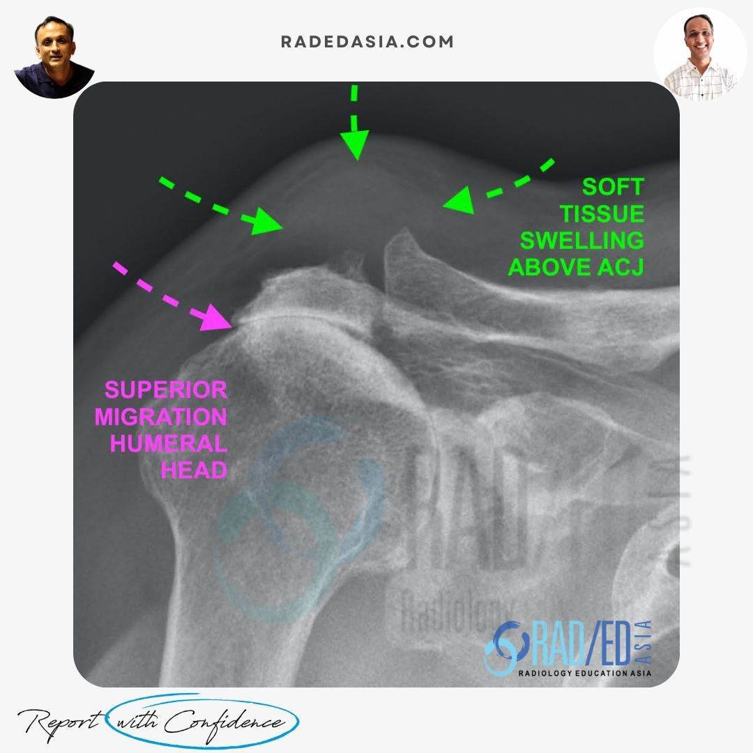
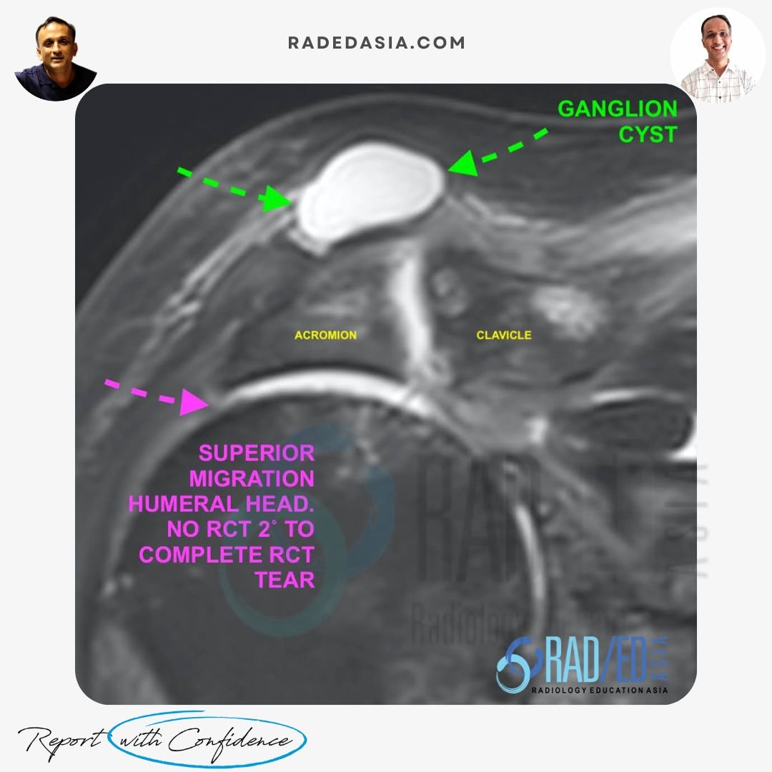
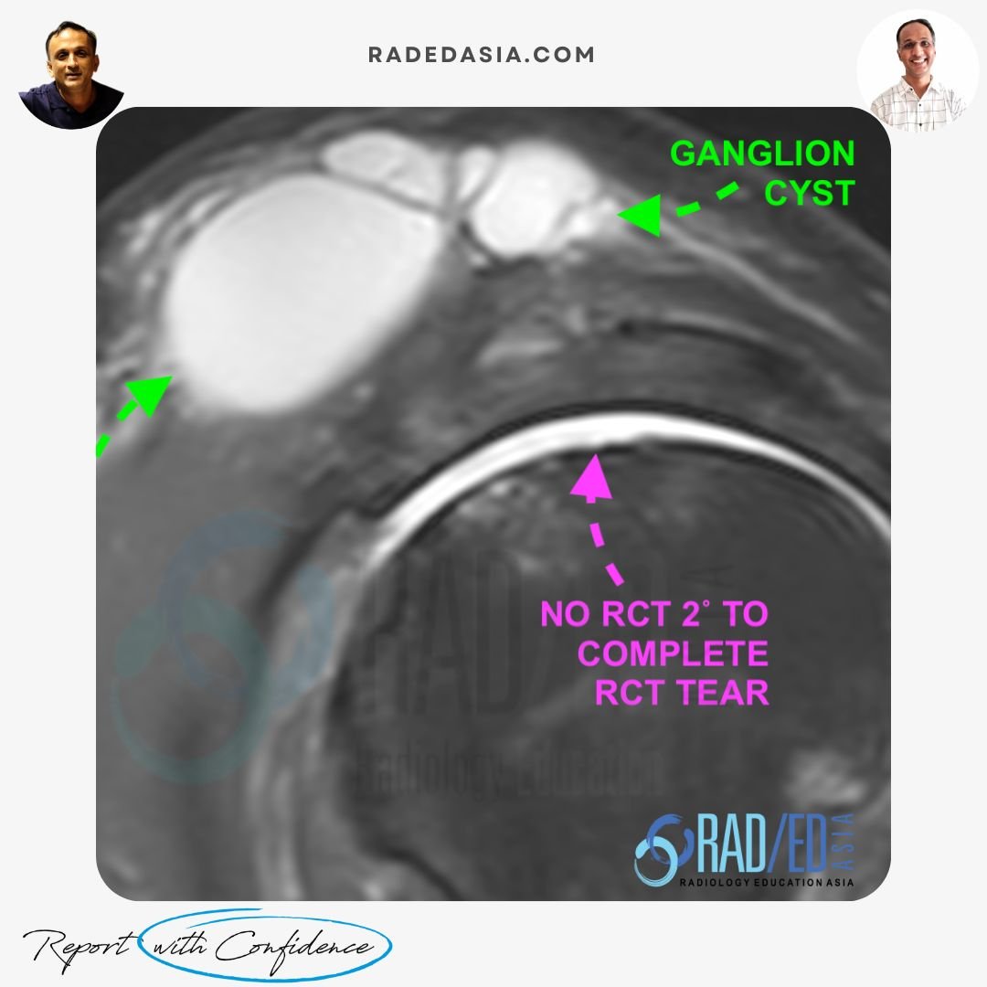
These cysts are associated with rotator cuff tears, usually complete tears. Complete RCT tears result in superior migration of the humeral head which erodes the ACJ capsule and fluid enters the ACJ from the Glenohumeral joint. There is also increased synovial fluid production in the ACJ. Eventually a tear in the superior margin of the ACJ capsule results in synovial fluid extension out of the AC joint, creating a ganglion cyst.![]()
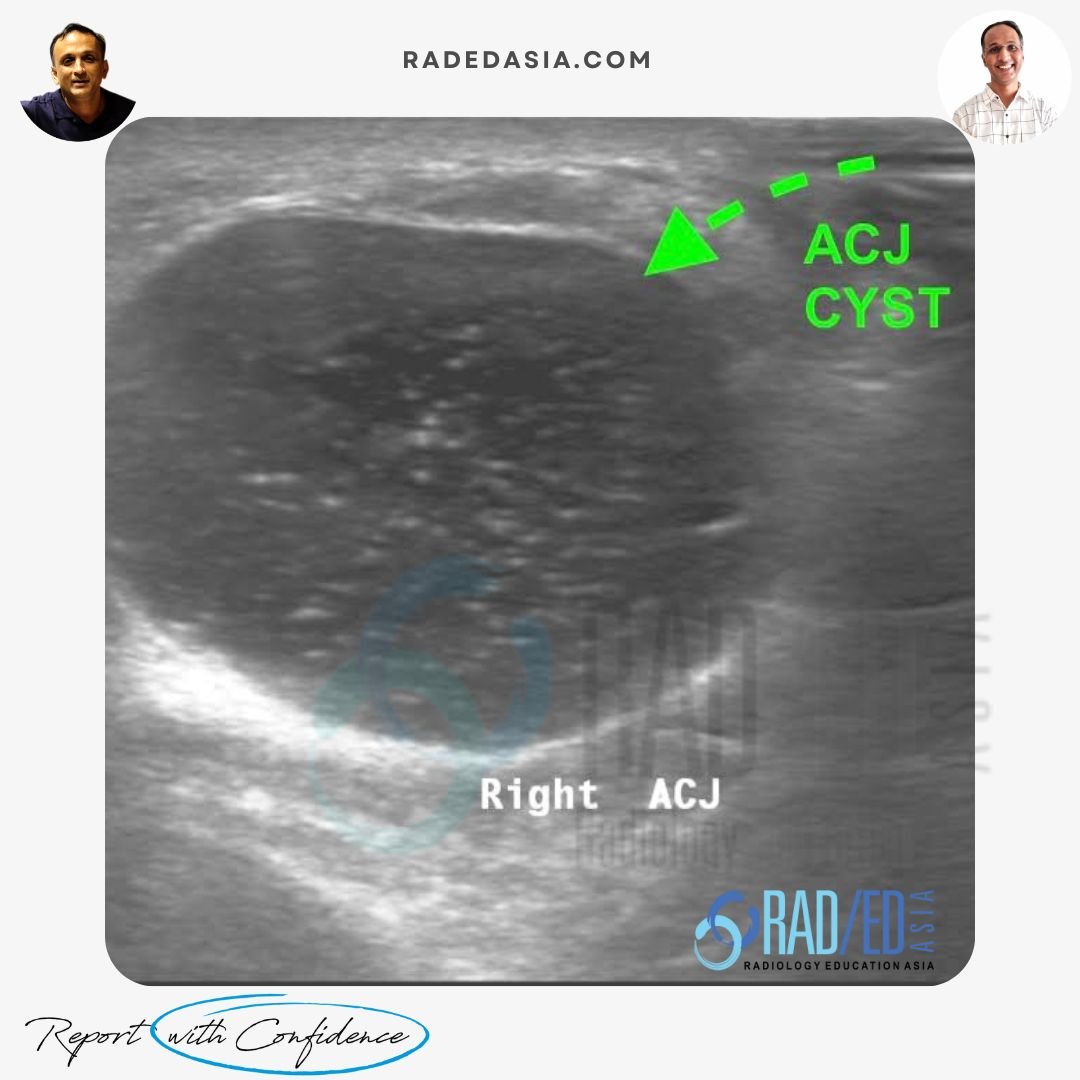
The Geyser sign is actually an arthrographic finding where contrast injected into the Glenohumeral joint/ACJ squirts out through a thin neck from the ACJ into the ganglion cyst. On MRI or ultrasound, you may or may not see the neck of the ACJ ganglion cyst.![]()
Whilst not technically difficult to aspirate and can be attempted, Type 2 cysts are likely to recur as there is a continuing communication between the GH joint, AC joint and the cyst. Surgical treatment is most commonly required for definitive treatment.![]()
![]()
Read Article: “Acromioclavicular Joint Cyst Formation Clinical Anatomy“, Read HERE![]()
Advanced AC joint arthritis. The rotator cuff is intact.![]()
Rotator cuff tears, usually complete tears.![]()
An arthrographic finding where contrast injected into the Glenohumeral joint or ACJ squirts out through a thin neck into the ganglion cyst.![]()
High recurrence rate due to the ongoing communication between the GH joint, AC joint, and the cyst. What are the limitations of US aspiration for Type 2 AC joint ganglion.![]()