arachnoiditis spine mri online radiology courses radedasia

ARACHNOIDITIS MRI SPINE ONLINE RADIOLOGY COURSE

Arachnoiditis Spine MRI Online Radiology Course

What are some of the common appearance of arachnoiditis on MRI?

Another three quick images with some basic key points from our online spine MRI courses.

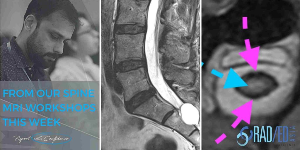
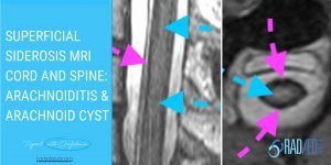
The distribution of nerve roots in the lumbar canal has to be symmetric. Any asymmetry indicates an abnormality.

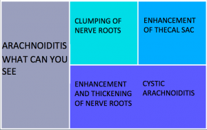
Clumping in arachnoiditis  occurs because fibrin lines the nerves post infection/ trauma/ bleeding which enables them to stick to each other.

The sac only appears empty as all the nerve roots are attached to the wall of the thecal sac.

We look at all of these topics in more detail in our SPINE MRI Mini Fellowships.

Click on the image below for more information.


  • Join our WhatsApp Group for regular educational posts. Message “JOIN GROUP” to +6594882623 (your name and number remain private and cannot be seen by others).

  • Get our weekly email with all our educational posts: https://bit.ly/whathappendthisweek

#radiology #radedasia #mri #spinemri #radiologyeducation #radiologycases #radiologist #radiologycme #radiologycpd #medicalimaging #imaging #radcme #rheumatology #arthritis #rheumatologist #orthopaedic #painphysician #chiropractic #chiropracter #physiotherapy #sportsmed #orthopaedic #ankylosingspondylitis #spondyloarthropathy #arachnoiditis #lumbar

#radedasia #mri #mskmri #radiología

Este sitio está destinado únicamente a profesionales médicos. El uso de este sitio se rige por nuestra Términos de servicio y Declaracion de privacidad que se puede encontrar haciendo clic en los enlaces. Acepte antes de continuar con el sitio web.
error: Clic derecho no disponible.