Weight-bearing x-ray showing Lisfranc joint instability with widening between the second metatarsal and cuneiform

Stay tuned on new Fellowships and learnings

WEIGHT-BEARING X-RAY: LISFRANC INSTABILITY – WHAT TO LOOK FOR (VIDEO)

WEIGHT BEARING X-RAY FOR LISFRANC INSTABILITY

Why weight-bearing views matter for Lisfranc joint injury

  • Early diagnosis of Lisfranc instability is crucial to prevent premature osteoarthritis in the mid foot.
  • Subtle instability patterns can be very difficult to identify on x-rays.
  • Plain radiographs may not effectively identify instability at the Lisfranc joint, especially on non-weight-bearing views.
  • Weight-bearing x-rays are important as they reveal subtle differences between the injured and uninjured sides.
  • However, weight bearing may not be possible in the acute phase due to pain.

If you find this helpful, please subscribe to the channel.

Assessing Instability on Weight-Bearing X-Ray

Look for lateral displacement of the second metatarsal in relation to the intermediate cuneiform on weight-bearing radiographs. A step-off of more than 3mm is abnormal.

Measure the distance between the medial cuneiform and the base of the second metatarsal on weight-bearing radiographs. A distance of more than 2.1mm is abnormal.

  • Comparison views of the contralateral foot are also very useful.
  • Do both non-weight-bearing and weight-bearing comparison views.
  • Subtle instability and widening can be easier to recognize when there is a direct comparison with the normal side.
Key Point
  • Weight-bearing radiographs provide greater reliability in diagnosing Lisfranc instability compared to non-weight-bearing views, revealing subtle differences between the injured and uninjured sides.
  • A C2–M2 step-off of more than 3mm between the second metatarsal and the intermediate cuneiform suggests instability.
  • A C1–M2 distance of more than 2.1mm between the medial cuneiform and the base of the second metatarsal suggests instability.
  • Comparison views of the contralateral foot, both weight-bearing and non-weight-bearing, help identify subtle instability.
  • Early diagnosis is important to prevent premature osteoarthritis developing in the mid foot.
Test Yourself on Some Common & Frequently Asked Questions

Plain radiographs may not effectively identify instability at the Lisfranc joint on non-weight-bearing views. Weight-bearing x-rays reveal subtle differences between the injured and uninjured sides that non-weight-bearing views can miss.

A step-off of more than 3mm between the second metatarsal and the intermediate cuneiform on weight-bearing radiographs.

A distance of more than 2.1mm between the medial cuneiform and the base of the second metatarsal on weight-bearing radiographs.

Subtle instability and widening can be easier to recognize when there is a direct comparison with the normal, uninjured side, ideally with both weight-bearing and non-weight-bearing views.

Early diagnosis is crucial to prevent premature osteoarthritis developing in the mid foot.

VIEW VIDEO: WEIGHT BEARING X-RAY LISFRANC INSTABILITY

If you wish to watch the video in language other than English, please view it on our YouTube Channel HERE

Our CPD & Learning Partners

Learn more about this condition & how best to report it in more detail in our Online Guided ANKLE or FOOT & TOE MSK MRI Mini Fellowship.

More by clicking on the images below

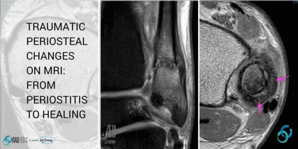
OUR OTHER POPULAR POSTS ON PERIOSTEALSTRIPPING: CLICK ON THE IMAGE BELOW

MRI periosteal stripping double black line cortex periosteum ankle shoulder trauma
TOBILLO
TOBILLO

Stay tuned on new Fellowships and learnings

error: Content is protected !!

Suscríbase a nuestro boletín

Esté atento a los nuevos lanzamientos y aprendizajes de las Fellowships

Fellowships

Mejor que las conferencias

Únase a nuestra comunidad de WhatsApp

Ayudamos a los radiólogos a aprender
haciendo e informa con confianza

Suscríbase a nuestro boletín

Esté atento a los nuevos lanzamientos y aprendizajes de las Fellowships

Fellowships

Mejor que las conferencias

Únase a nuestra comunidad de WhatsApp

Ayudamos a los radiólogos a aprender
haciendo e informa con confianza

Este sitio está destinado exclusivamente a las profesiones médicas. El uso de este sitio se rige por nuestras Condiciones de servicio y Declaración de privacidad, que puede consultar haciendo clic en los enlaces. Por favor, acéptelas antes de continuar en el sitio web.