STRESS FRACTURE FIBULA SHIN MRI RADIOLOGY XRAY
STRESS FRACTURE
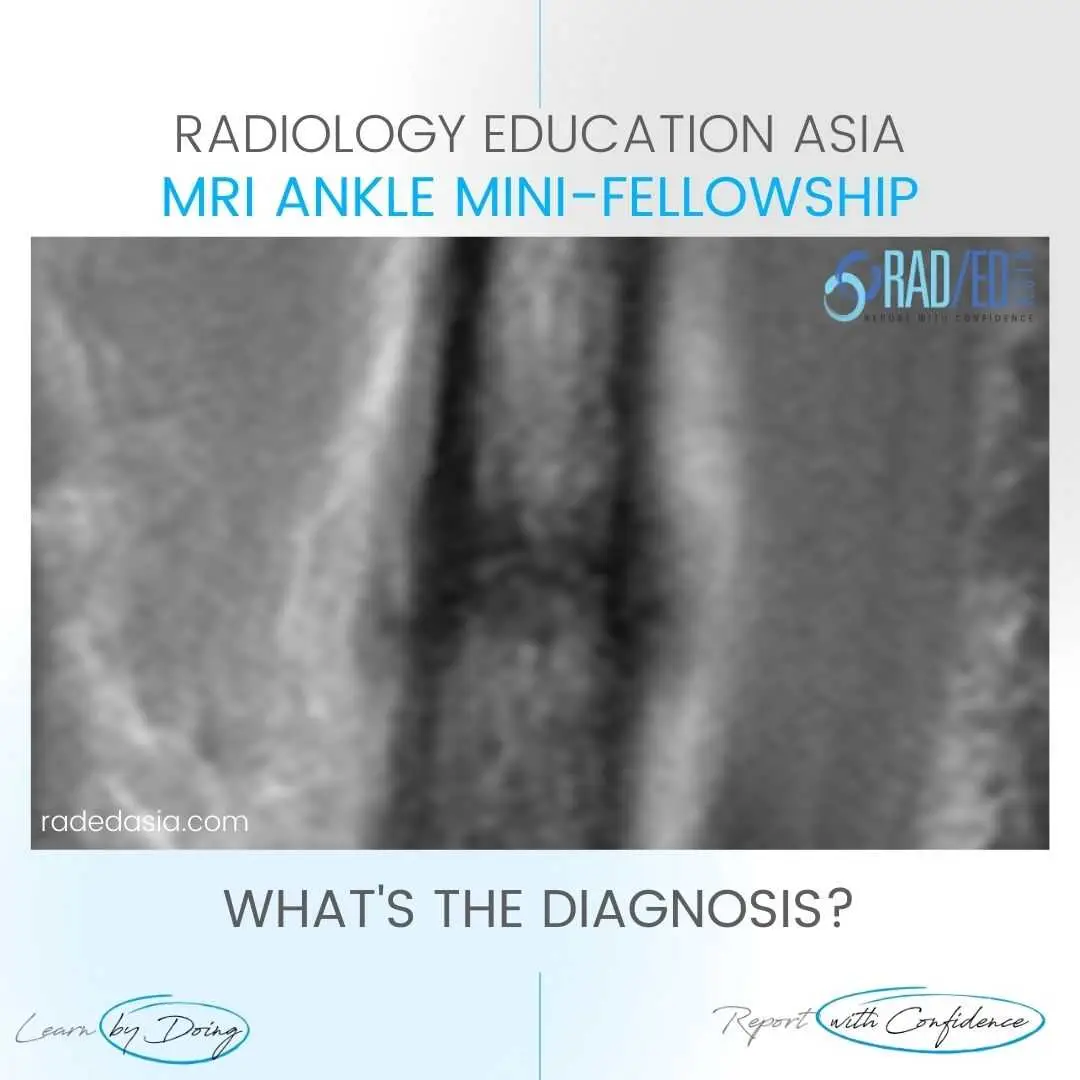
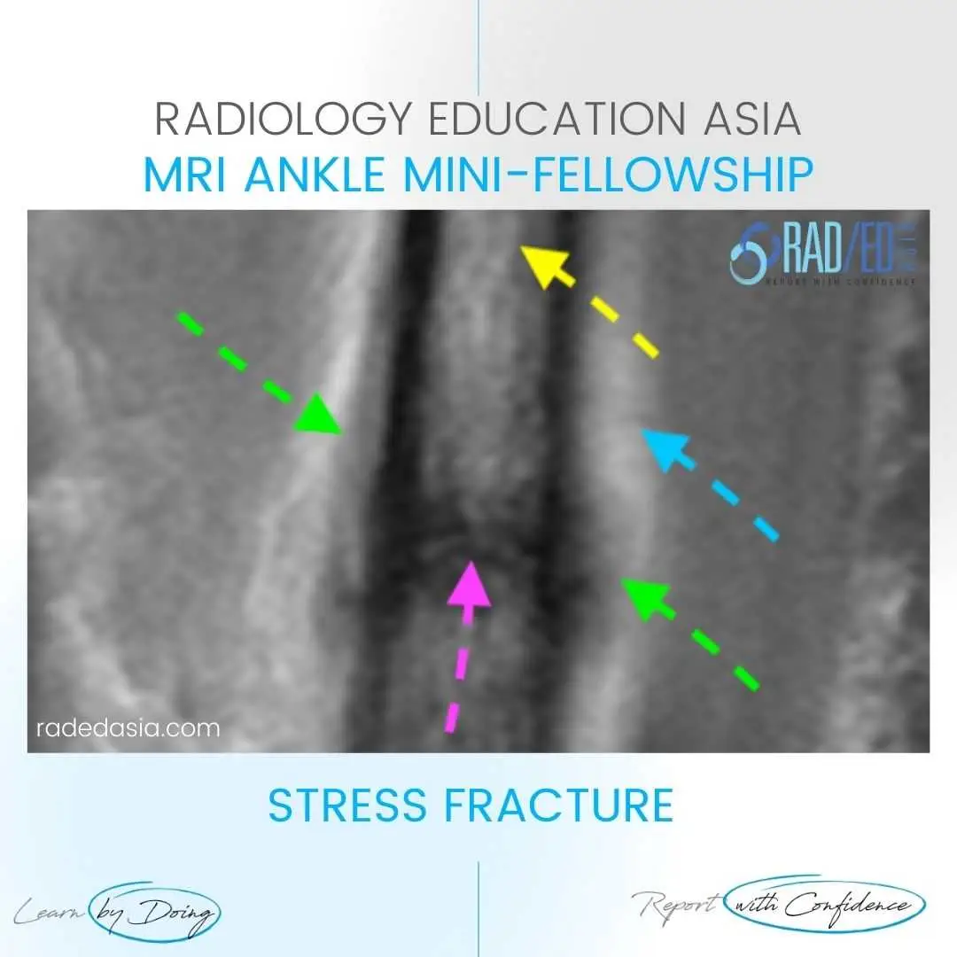
- Transverse stress fracture in the fibula shaft in a runner.
- This image demonstrates all the findings to look for in a stress fracture on MRI with.
- The Stress Fracture on MRI being the thin high signal line crossing transversely across the fibula (Pink arrow).
- The Low signal lines paralleling the cortex being Periosteal new bone formation (Green arrows).
- High Signal soft tissue changes being Soft tissue oedema and inflammation (Blue arrows).
- High Signal in the shaft being Bone marrow oedema (Yellow arrows).

- Thin high signal line crossing transversely across the fibula (Pink arrow).
- Periosteal new bone formation (Green arrows).
- Soft tissue oedema (Blue arrows).
- Bone marrow oedema (Yellow arrow).

#stressfracture #radiology #radedasia #mri #anklemri #msk #mriankle #mskmri #radiologyeducation #radiologycases #radiologist #radiologycme #radiologycpd #medicalimaging #imaging #radcme #rheumatology #arthritis #rheumatologist #sportsmed #orthopaedic #physio #physiotherapist

