hip labrum ossification mri

HIP LABRUM OSSIFICATION MRI: LABRAL ABNORMALITIES MORE THAN JUST TEARS PART 2

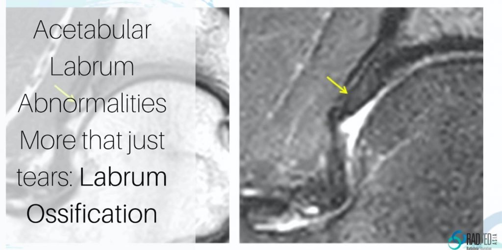
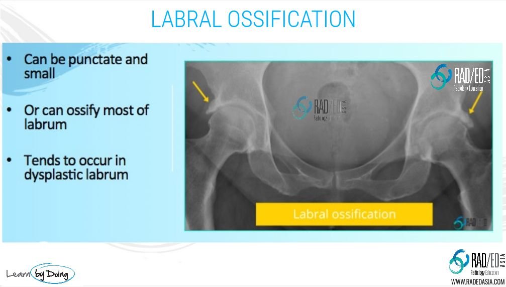
Acetabular labrum ossification is not common and I have been able to find only a few reports on it. The cases we have seen mostly have an underlying dysplastic, elongated labrum however there are reports of it occurring in a normal labrum. So what does it look like? Having learnt the hard way by missing them by looking only at the MRI, I find the best place to start is with the plain X-ray as it can be overlooked if assessing just the MRI.

hip-labrum-ossification-radedasia
 
Hip-labrum-ossifiction-xray-radedasia

Image 2 Above: Bilateral labral ossification in the same patient. Labrum is diffusely ossified. Ossification follows the shape of the labrum. MRI below.

hip-labrum-ossification-mri-radedasia

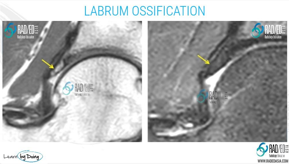
Image 3 Above: MRI for patient in Image 2. On the PD and PDFS scans ossification follows same signal as adjacent bone. Labrum is elongated and dysplastic.

hip-labrum-ossification-mri-rea-radedasia

Image 4 above: PD and PDFS ossification of the labrum centered on the labrum and follows labrum shape. Different patient to Image 2 and 3.

Learn more about HIP Imaging in our ONLINE
Guided MRI HIP Mini-Fellowship.


More by clicking on the images below.

#mrilabrum #labrumossification #labrummucoiddegeneration #radiology #radedasia #mri #hipmri #radiologyeducation #radiologycases #radiologist #radiologycme #radiologycpd #medicalimaging #imaging #radcme #rheumatology #arthritis #rheumatologist #orthopaedic #painphysician #podiatry #podiatrist #physiotherapy #sportsmed #orthopaedic #mskmri

#radedasia #mri #mskmri #radiología

Este sitio está destinado únicamente a profesionales médicos. El uso de este sitio se rige por nuestra Términos de servicio y Declaracion de privacidad que se puede encontrar haciendo clic en los enlaces. Acepte antes de continuar con el sitio web.
error: Clic derecho no disponible.