shoulder cartilage glenoid dislocation radiology education asia tuesday tip

MRI GLENOID CARTILAGE MRI SHOULDER WHERE & WHAT TO LOOK FOR SHOULDER DISLOCATIONS

GLENOID CARTILAGE MRI :What to look for and where to look for chondral damage in shoulder dislocations.


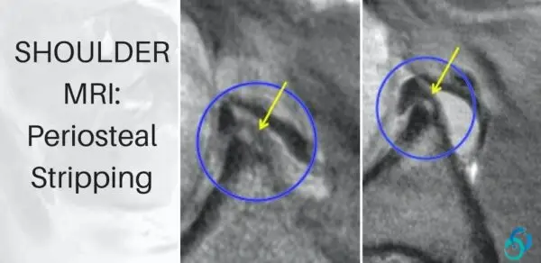
GLENOID CARTILAGE MRI: Glenoid cartilage damage can be seen with anterior and posterior shoulder dislocations on MRI and are part of the so-called GLAD lesions.  It’s important to first recognize the normal appearance of glenoid cartilage and its relationship to the labrum, which will help to understand where to look for cartilage damage in a shoulder dislocation.

The post looks at anterior dislocations but for posterior dislocations, it’s the same but reversed appearance.


radiology-education-asia-radedasia

Normal cartilage is intermediate grey (Blue arrows) signal on PD and PDFS.

  • Cartilage is firmly attached to the labrum (Pink arrows).
  • There should be no high signal between the intermediate grey of cartilage and the black signal of the labrum.
  • Look first at the chondro labral junction.
  • With a dislocation, the cartilage at the chondro labral junction is first damaged.
  • With chondral loss, the intermediate signal of cartilage (Blue Arrow) is replaced by high signal fluid (Orange arrow).

Our CPD & Learning Partners

error: Content is protected !!

Suscríbase a nuestro boletín

Esté atento a los nuevos lanzamientos y aprendizajes de las minibecas

Mini-becas

Mejor que las conferencias

Únase a nuestra comunidad de WhatsApp

Ayudamos a los radiólogos a aprender
haciendo e informar con confianza

Este sitio está destinado exclusivamente a las profesiones médicas. El uso de este sitio se rige por nuestras Condiciones de servicio y Declaración de privacidad, que puede consultar haciendo clic en los enlaces. Por favor, acéptelas antes de continuar en el sitio web.