Protegido: MRI Sacroileitis Chronic Changes
No hay extracto porque es una entrada protegida.
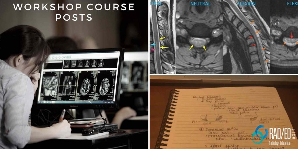
Arthritis Radiology Rheumatology Imaging
Imaging of Arthritis can be challenging because findings can be non specific and the clinical context of what we are seeing is very important in getting to or narrowing a diagnosis.
From 2021 all our posts on Arthritis Radiology and Rheumatology Imaging will be done in conjunction with Dr Joe Thomas a Senior Consultant Rheumatologist. who has a vast amount of clinical experience.
He also has a very strong interest in Imaging of Arthropathies and has joined us to bring a clinical perspective to the imaging and to advise on what rheumatologists want when we report their referrals.

ROMANUS LESION CORNER INFLAMMATORY LESION MRI CT The Romanus lesion (which is now called a Corner Inflammatory Lesion) is one of the earliest signs in the spine of inflammatory spondyloarthritis. It represents inflammatory changes at the insertion of the annulus of the disc to the endplate and is an enthesitis. It is more often seen …
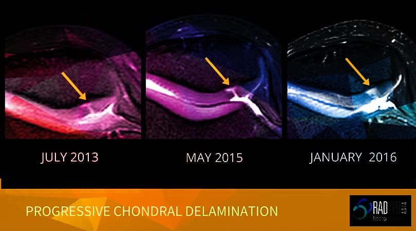
CHONDRAL DELAMINATION CARTILAGE MRI CHONDRAL DELAMINATION CARTILAGE MRI Chondral Delamination on MRI is not common but is seen often enough that we need to be aware of it and What to look for on MRI. OVERVIEW What is it? Chondral delamination is when cartilage separates and lifts off from its attachment with the cortex. …
CHONDRAL DELAMINATION CARTILAGE MRI: Quick Review Leer más »