SHOULDER DISLOCATION BONY BANKART GLENOID FRACTURE XRAY CT (VIDEO)
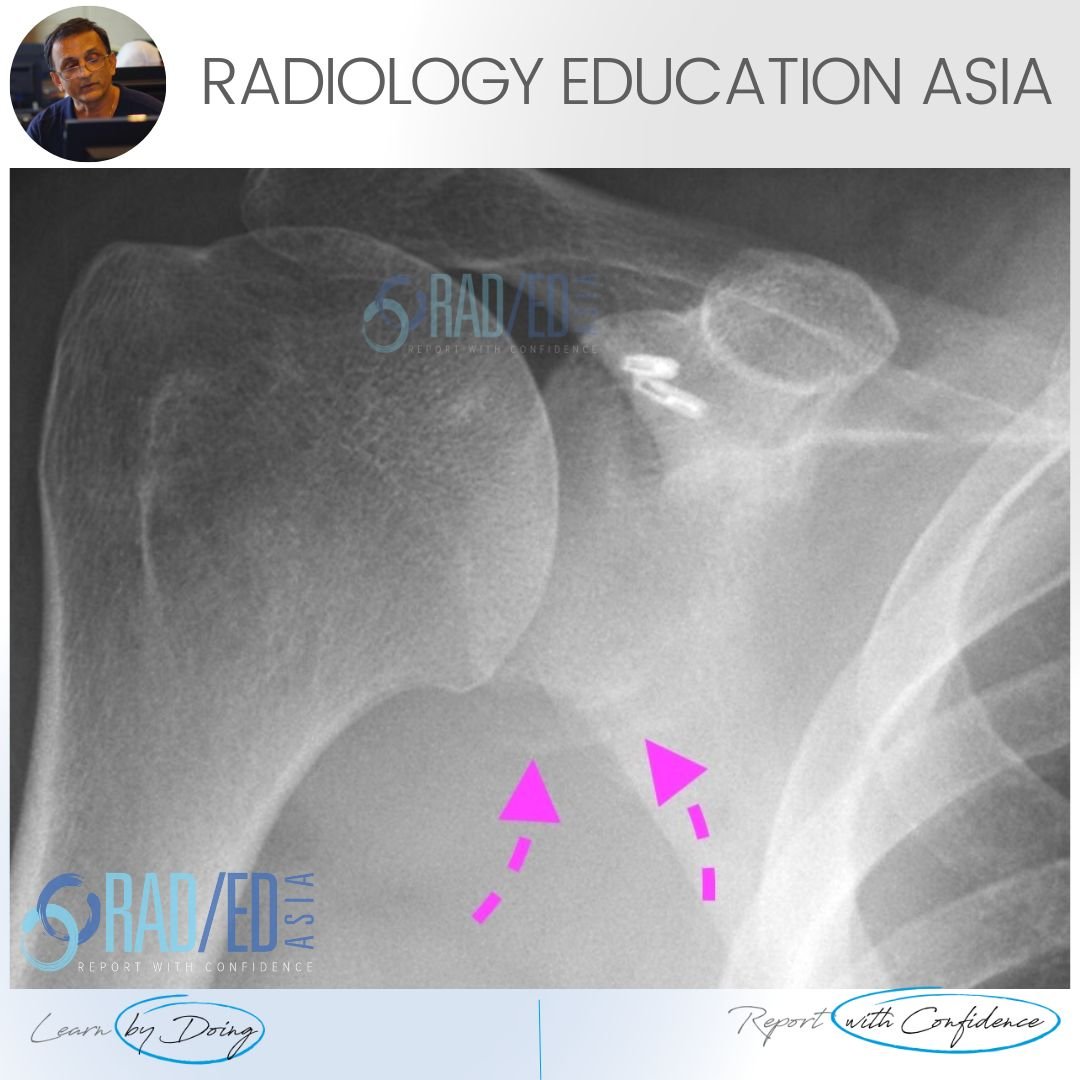
 Image Above: There is a thin fragment of bone at the inferior margin of the glenoid.

 Image Above: The inferior margin of the glenoid should normally be rounded and ovoid. Here it is flattened which is abnormal.

Bony Bankart lesions on x-ray can be very subtle. This is a larger bony bankart so its more obvious but sometimes all we see is a bit of bony irregularity or a suspicion of a fragment. If you are not sure, in the context of trauma I would always recommend a CT.
 Image Above: What we saw on the x-ray. Displaced bone fragment (Pink arrows) and flattening of the inferior margin of the glenoid (Green arrow).


