Denervation around the Shoulder: Quadrilateral Space Syndrome and Dislocation.
This post looks at denervation of the Axillary Nerve in Quadrilateral Space Syndrome and Axillary nerve damage post shoulder dislocation both of which are less common causes of denervation around the shoulder. They have a particular distribution so recognizing the muscles involved gives you the indication of the nerve involved.
![]()
Quadrilateral space is in the axillary region through which the axillary neurovascular bunde passes through.
Thought to be due to fibrous bands in the space compressing the neurovascular bundle in the quadrilateral space. Less commonly ganglia can cause compression.
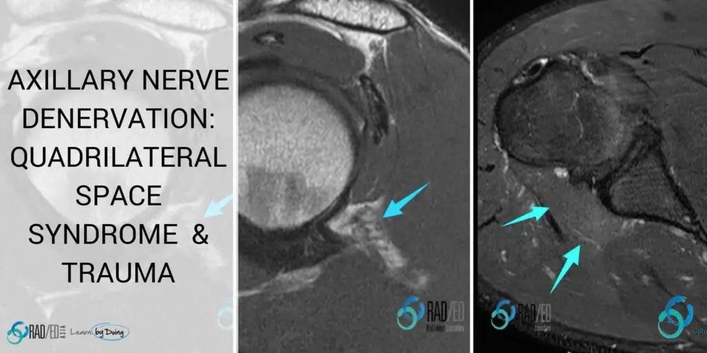
The main finding is atrophy and fatty infiltration isolated to the teres minor muscle. Less commonly the posterior fibres of deltoid can be involved.

Image Above: PD Coronal, Sagittal and Axial: Severe, isolated atrophy and fatty infiltration of teres minor (blue arrows).
Axillary nerve passes into the quadrilateral space and has two branches:
- Anterior division: Innervates deltoid mostly anterior and middle portions.
Posterior Division: Supplies teres minor and posterior portions of deltoid. Sensation to proximal lateral arm.
The axillary nerve is closely related to the inferior margin of the Glenohumeral joint and can be injured in an antero inferior dislocation. Most commonly its a neuropraxia with stretching of the nerve but this eventually recovers.
Look for denervation in muscles supplied by the axillary nerve (deltoid and teres minor) in the setting of a previous dislocation.
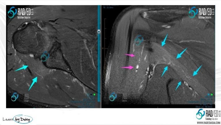 Image above: Post anterior dislocation changes of acute denervation (hyper-intensity on PDFS) in teres minor (Blue arrows) and posterior fibres of deltoid (Pink arrows). Distribution of changes indicates damage to posterior division of axillary nerve.
 Image above: Post anterior dislocation changes of acute denervation (hyper-intensity on PDFS) in teres minor (Blue arrows) and posterior fibres of deltoid (Pink arrows). Distribution of changes indicates damage to posterior division of axillary nerve.