ischiofemoral impingement radiology mri hip

Ischiofemoral impingement is a result of the quadratus femoris muscle being compressed between the ischial tuberosity and the posterior femur resulting initially in quadratus femoris oedema but can progress to tears and severe atrophy.

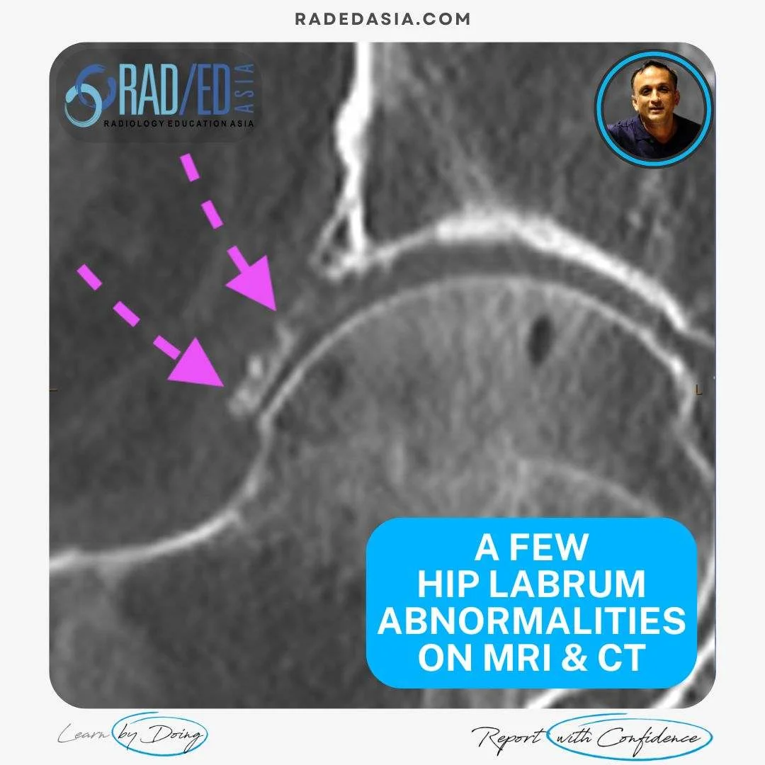
  • Initially there is oedema in the quadratus femoris muscle.


  • Look for Increased PDFS signal (Pink arrow) in the quadratus femoris muscle.


  • For the more detail and the progressive findings on MRI for Ischiofemoral impingement go to the related posts below and click on the link.

If your Browser is blocking the video, Please view it on our YouTube Channel HERE.

Learn more about HIP Imaging in our ONLINE 
Guided MRI HIP Mini-Fellowship.


More by clicking on the images below.

hip labrum tear paralabral cyst mucoid degeneration ossification mri ct radedasia
HIP
pyriformis syndrome pudendal nerve quadratus femoris ischiofemoral impingement ultrasound scan inject radedasia
HIP

>#radiology #radedasia #mri #hipmri #msk #mrihip #mskmri #radiologyeducation #radiologycases #radiologist #rads #radiologystudent #radiologycme #radiologycpd #medicalimaging #imaging #radcme #rheumatology #arthritis #rheumatologist #sportsmed #orthopaedic #physio #physiotherapist #ischiofemoralimpingement

#radedasia #mri #mskmri #radiología

Este sitio está destinado únicamente a profesionales médicos. El uso de este sitio se rige por nuestra Términos de servicio y Declaracion de privacidad que se puede encontrar haciendo clic en los enlaces. Acepte antes de continuar con el sitio web.
error: Clic derecho no disponible.