mri shoulder denervation parsonage turner syndrome supraspinatus infraspinatus paralabral cyst suprascapular spinoglenoid notch anatomy radiology education asia

MRI SHOULDER: PARALABRAL CYSTS, DENERVATION AND PARSONAGE TURNER SYNDROME

DENERVATION: What are the causes & What does it look like:


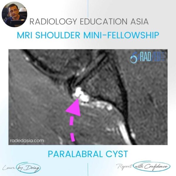
On MRI muscles around the shoulder can show features of denervation. So what are the causes and what does it  look like. Most commonly its due to nerve compression from a paralabral cyst secondary to a labral tear (usually a SLAP tear).  It can also be due to brachial neuritis (Parsonage Turner Syndrome) with no evidence of a compressing mass. In this post we look at paralabral cysts and in the next post at Parsonage Turner Syndrome.


ANATOMY:

The suprascapular nerve supplies the supra and infraspinatus muscles. The main branch runs in the suprascapular notch and divides into two branches one supplies supraspinatus and the second exits via the spinoglenoid notch and runs in the groove inferior to the notch and supplies infraspinatus.


mri-shoulder-denervation-parsonage-turner-syndrome-infraspinatus-supraspinatus-paralabral-cyst-spinoglenoid-suprascapular-notch-radiology-education-asia-radedasia

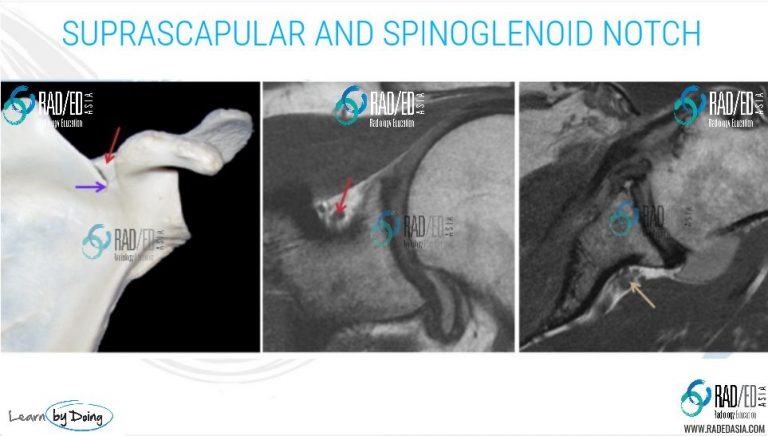
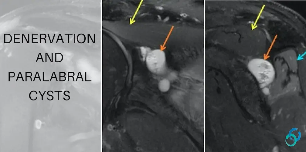
Image Above: Red arrow suprascapular notch. Purple arrow spinoglenoid notch. Gold arrow inferior to spinoglenoid notch  where branch to infraspinatus runs.

mri shoulder denervation parsonage turner syndrome infraspinatus supraspinatus paralabral cyst radiology education asia

mri-shoulder-denervation-parsonage-turner-syndrome-infraspinatus-supraspinatus-paralabral-cyst-suprascapular-notch-radiology-education-asia-radedasia

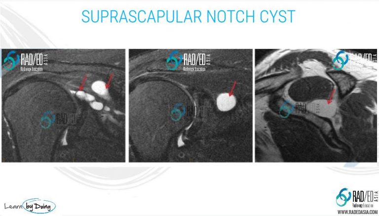
Image Above: Multiloculated cyst (red arrow) lying in the suprascapular notch where it could potentially compress the suprascapular nerve. No evidence of denervation, muscle signal normal.

mri-shoulder-denervation-parsonage-turner-syndrome-infraspinatus-supraspinatus-paralabral-cyst-spinoglenoid-suprascapular-radiology-education-asia-radedasia

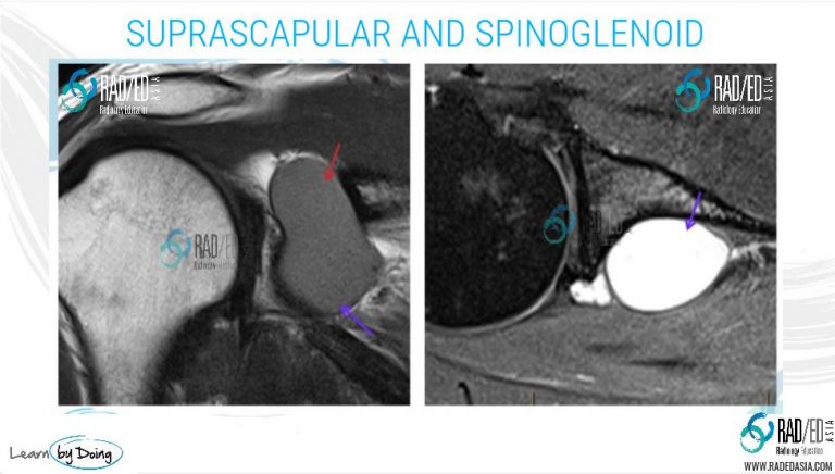
Image Above: Cyst in the suprascapular (red arrow) and spinoglenoid notch extending to the groove inferior to the notch (purple arrow). No evidence of denervation.

mri-shoulder-denervation-parsonage-turner-syndrome-infraspinatus-supraspinatus-paralabral-cyst-suprascapular-spinoglenoid-notch-anatomy-radiology-education-asia-radedasia

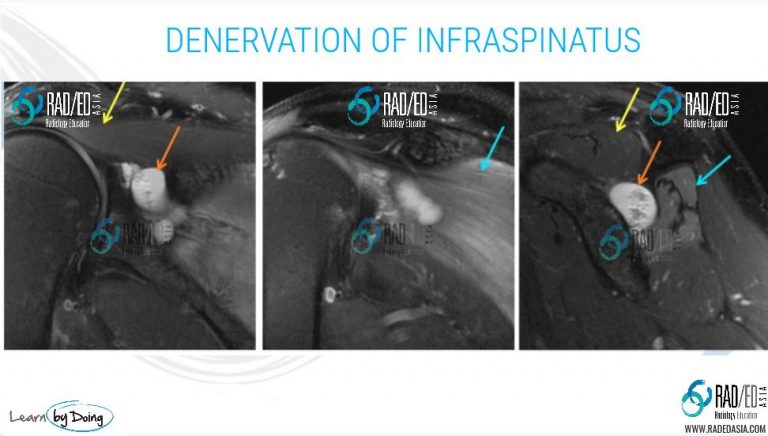
Image Above: Suprascapular and spinoglenoid  notch cyst (orange arrow) with denervation of infraspinatus (orange arrow) but no denervation of supraspinatus (yellow arrow). Acute denervation as infraspinatus is high signal on PDFS with no evidence of volume loss of the muscle. fatty replacement of the muscle would be better assessed on a non fat sat PD or T1. 

mri-shoulder-denervation-parsonage-turner-syndrome-supraspinatus-infraspinatus-paralabral-cyst-suprascapular-spinoglenoid-notch-radiology-education-asia-radedasia

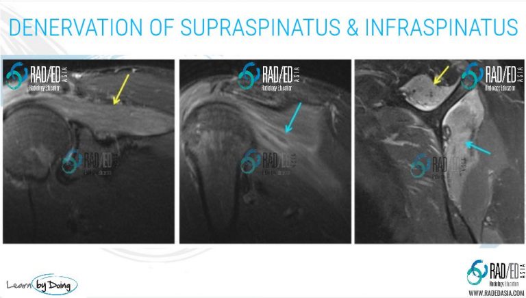
Image Above: Denervation of both supraspinatus (yellow arrow) and infraspinatus (blue arrow).


RELATED POSTS THAT INCREASE YOUR DEPTH OF KNOWLEDGE ON THIS TOPIC:

axillary-nerve-denervation-quadrilateral-space-syndroma-dislocation-radiology-education-asia-radedasia

denervation lumbar mri paraspinal

Learn more at our Online and Onsite GUIDED LEARNING mini-fellowships. Click on the image below:

online radiology conference course mri fellowship

What’s the Purpose of the Tuesday Tips? Many of the Tuesday Tip posts arise from questions or difficulties that people bring up in the Mini Fellowships. These posts are meant to be short and concise to answer those questions.


#radedasia

#radedasia #mri #mskmri #radiología

Este sitio está destinado únicamente a profesionales médicos. El uso de este sitio se rige por nuestra Términos de servicio y Declaracion de privacidad que se puede encontrar haciendo clic en los enlaces. Acepte antes de continuar con el sitio web.
error: Clic derecho no disponible.