RETROCALCANEAL BURSITIS RADIOLOGY MRI
- RETROCALCANEAL BURSITIS.
- ACHILLES TENDONOSIS.
- The retrocalcaneal bursa lies anterior and deep to the Achilles tendon postero superior to the calcaneum.
- The normal bursa has minimal fluid so on MRI you may see nothing or just a thin amount of fluid without synovial thickening.
- For retrocalcaneal bursitis on MRI look for contained fluid signal intensity in the expected location of the bursa.
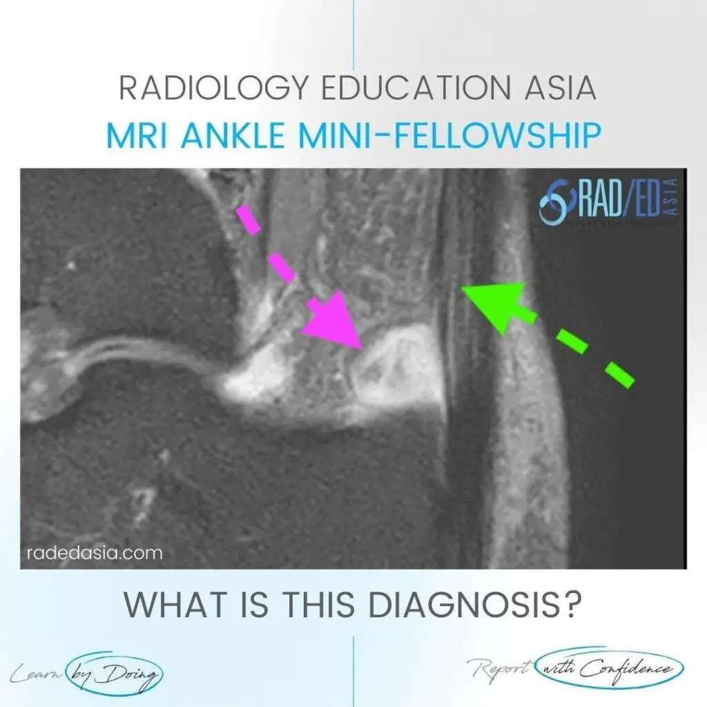
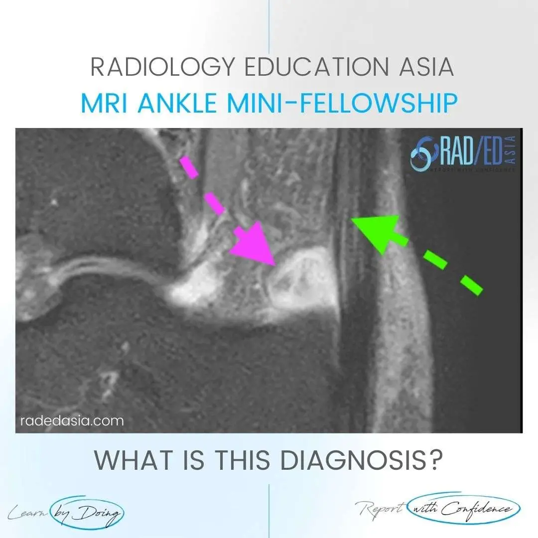
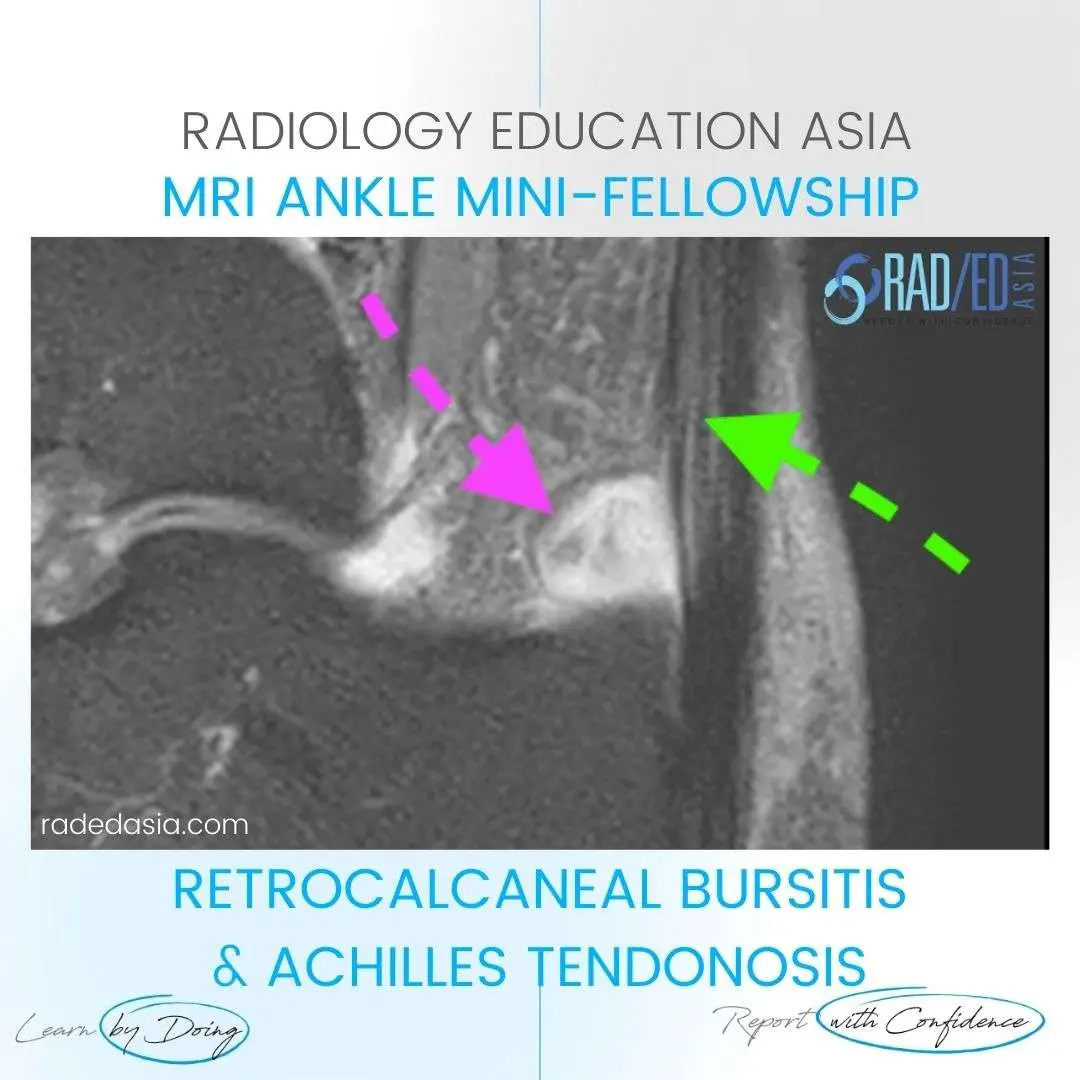
- In this case there is significant fluid in the bursa (Pink arrow) in keeping with bursitis.
- The Achilles tendon is usually abnormal with tendonosis or tears. In this case, increased intermediate signal and expansion Achilles tendon (Green Arrow) in keeping with tendonosis.
- Fluid present anterior to the Achilles tendon ( Pink arrow).
- Increased intermediate signal and expansion Achilles tendon ( Green Arrow).
#radiology #radedasia #mri #anklemri #msk #mriankle #mskmri #radiologyeducation #radiologycases #radiologist #radiologystudent #radiologycme #radiologycpd #medicalimaging #imaging #radcme #rheumatology #arthritis #rheumatologist #sportsmed #orthopaedic #physio #physiotherapist