Este sitio está destinado exclusivamente a las profesiones médicas. El uso de este sitio se rige por nuestras Condiciones de servicio y Declaración de privacidad, que puede consultar haciendo clic en los enlaces. Por favor, acéptelas antes de continuar en el sitio web.

CHONDRAL MRI KNEE: WHAT’S NORMAL & WHY (VIDEO)
CARTILAGE CHONDRAL MRI: Why normal cartilage has a laminar type appearance
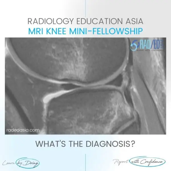
To understand abnormal cartilage on MRI, in say in a MRI of the knee with osteoarthritis, its important to have an understanding of the normal MRI appearance of cartilage.
So if we want to understand cartilage degeneration we need to first know what normal cartilage looks like, which will help to make a more accurate diagnosis.
Normal cartilage on MRI has a tri-laminar appearance, why is that?
In today’s video, we look at
- Normal cartilage on knee MRI as an example of MRI appearance of cartilage in other joints
- The variations in appearance of normal cartilage on MRI and
- A brief look at some theories of why there are different signal intensities in normal cartilage.
If you find the video helpful, please subscribe to the channel.
If your Browser is blocking the video, Please Click HERE to view it on our YouTube channel.
If you find the video helpful, please subscribe to the channel.
- Join our WhatsApp RadEdAsia community for regular educational posts at this link: https://bit.ly/radedasiacommunity
- Get our weekly email with all our educational posts: https://bit.ly/whathappendthisweek


