KNEE

LEARN MSK MRI KNEE ONLINE     knee icon radedasia

KNEE MSK MRI RADIOLOGY: We regularly post on Knee MRI and our Knee MRI Courses with all the posts on this page.

Knee MRI can be difficult due to the complex anatomy. In our posts we have concise explanation of the Knee MRI anatomy, the macroscopic pathology that’s relevant to the Imaging and the Imaging findings in various pathologies.

Learn where to Look, What to Look for and How to Report Knee MRI More Confidently

learn msk mri online knee mri cartilage surface lesions radedasia

CARTILAGE DAMAGE MRI KNEE : SURFACE LESIONS (VIDEO)

CARTILAGE MRI KNEE LESIONS: CHONDRAL DAMAGE KNEE Cartilage MRI Chondral Damage: What are the Cartilage surface lesions to report   Cartilage MRI : The 3 simple MRI appearances of surface cartilage damage and injury. Cartilage Fibrillation, Cartilage Fissure and Chondral Delamination How to easily find and report them on MRI.   MRI cartilage early damage. …

CARTILAGE DAMAGE MRI KNEE : SURFACE LESIONS (VIDEO) Read More »

chondral mri learn msk mri online knee mri cartilage normal radedasia

CHONDRAL MRI KNEE: WHAT’S NORMAL & WHY (VIDEO)

NORMAL CARTILAGE CHONDRAL MRI KNEE CARTILAGE CHONDRAL MRI: Why normal cartilage has a laminar type appearance To understand abnormal cartilage on MRI, in say in a MRI of the knee with osteoarthritis, its important to have an understanding of the normal MRI appearance of cartilage.    So if we want to understand cartilage degeneration we …

CHONDRAL MRI KNEE: WHAT’S NORMAL & WHY (VIDEO) Read More »

proximal tibio fibular joint mri normal anatomy ligaments biceps femoris online radiology courses conference education asia

TUESDAY TIP: MRI OF THE PROXIMAL TIBIOFIBULAR JOINT (PART 2)

Today’s Tuesday Tip continues with the Proximal TibioFibular Joint. On MRI at the Proximal TibioFibular Joint ( PTFJ) one thing that can be confusing is the tibial attachment of the Biceps Femoris Tendon which can be mistaken for the AnteroSuperior TibioFibular Ligament. The Biceps Femoris tendon attaches predominantly to the fibula head but there is …

TUESDAY TIP: MRI OF THE PROXIMAL TIBIOFIBULAR JOINT (PART 2) Read More »

arcuate segond fracture fibular styloid radiology education asia tuesday tip

ARCUATE FRACTURE KNEE X-RAY, SEGOND FRACTURE, LATERAL KNEE FRACTURES

ARCUATE FRACTURE, SEGOND FRACTURE, LATERAL KNEE FRACTURES ARCUATE FRACTURE KNEE X-RAY , SEGOND FRACTURE, LATERAL FIBULAR FRACTURE This post looks at the 3 fractures (Segond Fracture, Arcuate Fracture and Lateral Fibular Fracture ) that can be seen on the lateral aspect of the knee x-ray which are indicative of underlying tendon or ligament involvement. The …

ARCUATE FRACTURE KNEE X-RAY, SEGOND FRACTURE, LATERAL KNEE FRACTURES Read More »

knee mcl pellegrini steida mri xray radiology education asia radedasia

TUESDAY TIP: Pellegrini Stieda Lesion and Syndrome What to Look for on MRI and Xray

  Good Evening today’s Tuesday Tip is on What to look for on Xray and MRI in Pellegrini Stieda Lesions. The Pellegrini Stieda Syndrome is when there is associated pain and movement restriction and is a combination of the imaging and clinical findings. More in the image above. Whats the Purpose of the Tuesday Tips?: …

TUESDAY TIP: Pellegrini Stieda Lesion and Syndrome What to Look for on MRI and Xray Read More »

mri-cartilage-early-changes-delamination-radedaia-radiology-education-asia

MRI CARTILAGE CHONDRAL DAMAGE: CARTILAGE DELAMINATION & EARLY CARTILAGE CHANGES

MRI CARTILAGE CHONDRAL DAMAGE MRI CARTILAGE CHONDRAL DAMAGE & LESIONS: What to look for and How to report. What are the early changes of chondral damage we should see and report. This post covers the 3 Things to look for. MRI CARTILAGE CHONDRAL DAMAGE: 3 THINGS TO LOOK FOR MRI CARTILAGE FIBRILLATION Surface irregularity of …

MRI CARTILAGE CHONDRAL DAMAGE: CARTILAGE DELAMINATION & EARLY CARTILAGE CHANGES Read More »

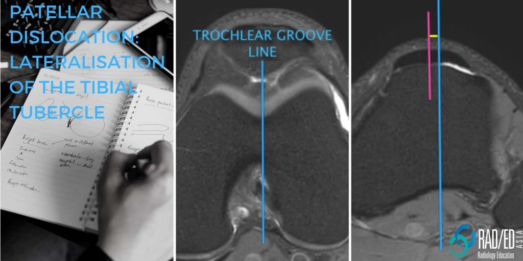
patellar dislocation lateralisation tibial tubercle radiology education asia

MRI TIBIAL TUBERCLE HOW TO ASSESS LATERALISATION OF THE TIBIAL TUBEROSITY

MRI TIBIAL TUBERCLE MRI TIBIAL TUBERCLE : HOW TO ASSESS LATERALISATION OF THE TIBIAL TUBERCLE   Lateralization of the tibial tubercle is one of the predisposing factors for Patellar Dislocation.    In this post we look at what the normal position is, how to measure lateralization and what the differences between CT and MRI measurements …

MRI TIBIAL TUBERCLE HOW TO ASSESS LATERALISATION OF THE TIBIAL TUBEROSITY Read More »

This site is intended for Medical Professionals only. Use of this site is governed by our Terms of Service and Privacy Statement which can be found by clicking on the links. Please accept before proceeding to the website.
error: Right Click not available.