mucoid-degeneration-hip-labrum

HIP ACETABULAR LABRUM MRI MORE THAN JUST TEARS: MUCOID DEGENERATION

There is a lot written about acetabular labral tears but there are other abnormalities of the labrum that can be precursors to or associated with tears. Here we look at Mucoid Degeneration.

Mucoid degeneration of the acetabular labrum is not uncommon, but Ive not been able to find too much written about it apart from passing references to it in articles dealing with the labrum.

MRI MUCOID DEGENERATION: SOME BACKGROUND

  1. The MRI findings are T2/PD hyper-intensity and enlargement of the labrum which can be focal or diffuse.
  2. Intra labral cysts can also develop.
  3. We see it more often in the anterosuperior and superolateral labrum
  4. The MRI changes are similar to mucoid degeneration of other structures like the ACL or degeneration that can occur in a discoid meniscus.
  5. Mucoid degeneration can occur in an otherwise normal labrum or in dysplastic labrums associated with acetabular dysplasia.

acetabular-labrum-hip-mucoid-degeneration-radedasia

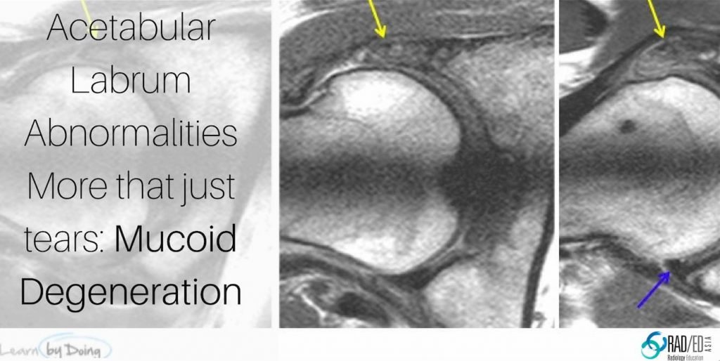
Image Above: PD and PDFS scans. Enlargement and diffuse hyper-intensity superolateral labrum (yellow arrow). Signal intensity is high but not as high as fluid.

hup-labrum-mucoid-degeneration-cystic-change-radedasia

Image above: PDFS all same patient: Early cystic change (yellow arrow) within mucoid degeneration. More posterior image demonstrates mucoid degeneration (blue arrows) without cystic change where the signal is increased but not approximating fluid signal.

hip-labrum-tear-mucoid-degeneration-radedasia

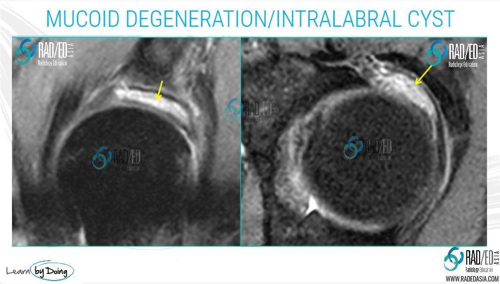
Image above: PDFS: Areas of hyper-intensity (yellow arrow) approximating fluid signal suggesting early cyst formation.

hip-labrum-mucoid-degeneration-labral-tear-radiology-education-radedasia

Image above all from the same patient: First image (Red arrow) early changes of mucoid degeneration with slight increased signal. Second image (Yellow arrow) more pronounced changes. Third image (blue arrow) associated labral tear at margins of mucoid degeneration.


labrum-hip-mucoid-localized-radedasia

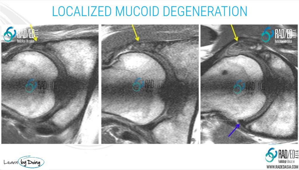
Image above: PD, Radial scan. Localized progressive increase in mucoid degeneration anterior labrum (yellow arrow) and a completely normal posterior labrum (blue arrow).


mucoid-degeneration-hip-labrum-extensive-radedasia

Image Above: Generalized cystic mucoid degeneration (yellow arrows). Only the postero inferior labrum was spared (not shown). Green lines in 2nd image reflect the level of the first image axial scan. Blue arrow first image is a paralabral cyst from a labral tear.

Learn more about HIP Imaging in our ONLINE
Guided MRI HIP Mini-Fellowship.


More by clicking on the images below.

#mrilabrum #labrumossification #labrummucoiddegeneration #radiology #radedasia #mri #hipmri #radiologyeducation #radiologycases #radiologist #radiologycme #radiologycpd #medicalimaging #imaging #radcme #rheumatology #arthritis #rheumatologist #orthopaedic #painphysician #podiatry #podiatrist #physiotherapy #sportsmed #orthopaedic #mskmri

#radedasia #mri #mskmri #radiology

This site is intended for Medical Professionals only. Use of this site is governed by our Terms of Service and Privacy Statement which can be found by clicking on the links. Please accept before proceeding to the website.
error: Right Click not available.