SHOULDER

LEARN MSK MRI SHOULDER ONLINEshoulder icon radedasia


SHOULDER MSK MRI RADIOLOGY: We regularly post on Shoulder MRI and our Shoulder MRI Courses with all the posts on this page.


Shoulder MRI can be difficult due to the complex anatomy. In our posts we have concise explanation of the Shoulder MRI anatomy, the macroscopic pathology that’s relevant to the Imaging and the Imaging findings in various pathologies.


Learn where to Look, What to Look for and How to Report Shoulder MRI More Confidently

rotator cuff tendinitis mri supraspinatus tendinosis bursitis radiology

ROTATOR CUFF SUPRASPINATUS TENDINOSIS TENDINITIS MRI: WHAT’S WRONG WITH THE SST?

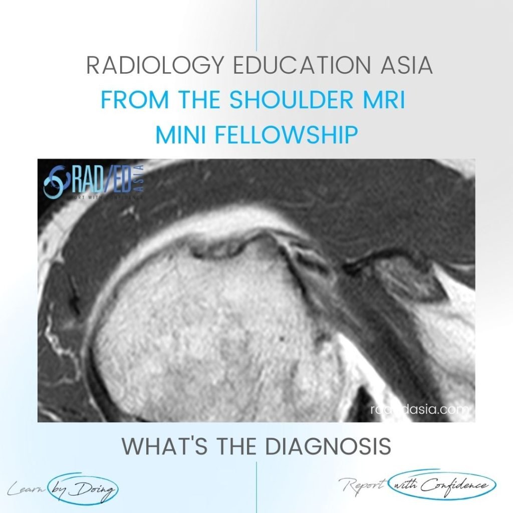
MOVE SLIDER ICON (ARROWS) LEFT TO SEE THE DIAGNOSIS WHAT ARE THE FINDINGS Diffuse Increased signal (intermediate) in the Supraspinatus Tendon. WHAT'S THE Dx SUPRASPINATUS TENDINITIS/TENDINOSIS.  There is diffuse increased signal in the supraspinatus tendon (Pink arrows). The signal increase is intermediate which is seen on tendinitis and not fluid type signal which is seen …

ROTATOR CUFF SUPRASPINATUS TENDINOSIS TENDINITIS MRI: WHAT’S WRONG WITH THE SST? Read More »

shoulder labrum normal variant buford complex sublabral foramen hole mri

SHOULDER LABRUM NORMAL VARIANTS: WHICH VARIANTS OCCUR HERE (VIDEO)

SHOULDER LABRUM NORMAL VARIANTS DISCUSSION Sublabral Foramen & Buford Complex. FINDINGS Two normal variants occur in superior portion of the anterior labrum: Sublabral Foramen (also called Sublabral Hole), The Buford complex. SHOULDER LABRUM NORMAL VARIANTS VIDEO If your Browser is blocking the video, Please view it on our YouTube Channel HERE. Learn more about SHOULDER …

SHOULDER LABRUM NORMAL VARIANTS: WHICH VARIANTS OCCUR HERE (VIDEO) Read More »

learn shoulder mri biseps disloc cyst radedasia

BICEPS DISLOCATION MRI SHOULDER RADIOLOGY

BICEPS DISLOCATION MRI SHOULDER RADIOLOGY DISCUSSION ON BICEPS DISLOCATION Extra articular Biceps dislocation. This is an Extra articular Biceps dislocation into a partial tear the Subscapularis tendon. FINDINGS IN BICEPS DISLOCATION Empty Bicipital Groove (Blue arrow).   Biceps tendon dislocated (Pink arrow).   Partial tear of Subscapularis tendon (Green arrow). High signal is seen within the …

BICEPS DISLOCATION MRI SHOULDER RADIOLOGY Read More »

mri shoulder ligaments mghl middle glenohumeral ligament radedasia

SHOULDER MRI MIDDLE GLENO HUMERAL LIGAMENT

MIDDLE GLENO HUMERAL LIGAMENT MRI SHOULDER MGHL DISCUSSION ON MGHL This is the normal MGHL (Middle Gleno Humeral Ligament). It lies adjacent and anterior to the labrum (Green arrow) and extends inferiorly to join the anterior capsule (Blue arrow) deep to subscapularis. FINDINGS IN MGHL Well defined low signal structure (Pink arrows) adjacent to the …

SHOULDER MRI MIDDLE GLENO HUMERAL LIGAMENT Read More »

mri shoulder labrum normal variation tear radedasia

MRI SHOULDER LABRUM CARTILAGE UNDERCUTTING: How is it different to Normal and Tears.

MRI SHOULDER LABRUM CARTILAGE UNDERCUTTING : WHATS NORMAL, VARIATION AND TEAR On MRI, Shoulder Undercutting by cartilage of the labrum can result in the appearance of a tear. How do you diagnose this? HOW TO REPORT IT NORMAL SHOULDER LABRUM & CARTILAGE ATTACHMENT A normal MRI of the Shoulder usually demonstrates the labrum firmly attached …

MRI SHOULDER LABRUM CARTILAGE UNDERCUTTING: How is it different to Normal and Tears. Read More »

philippines radiology conference ctmrisp mri msk mri online shoulder

CTMRISP SHOULDER MSK MRI COURSE ONLINE RADIOLOGY MAY 2024

Home CTMRISP MRI SHOULDER MINI FELLOWSHIP COURSE BEGINS 4th MAY 2024 We are very happy to be collaborating with CTMRISP to have this MRI Shoulder Online Course specifically for CTMRISP Fellows in Training and CTMRISP Fellows. Our new and updated 2024 MSK MRI Course Online in MRI SHOULDER  will cover the common abnormalities we see …

CTMRISP SHOULDER MSK MRI COURSE ONLINE RADIOLOGY MAY 2024 Read More »

This site is intended for Medical Professionals only. Use of this site is governed by our Terms of Service and Privacy Statement which can be found by clicking on the links. Please accept before proceeding to the website.
error: Right Click not available.