Dx

spine ligaments mri injury radiology interspinous trauma

SPINE LIGAMENTS MRI INJURY RADIOLOGY TRAUMA INTERSPINOUS

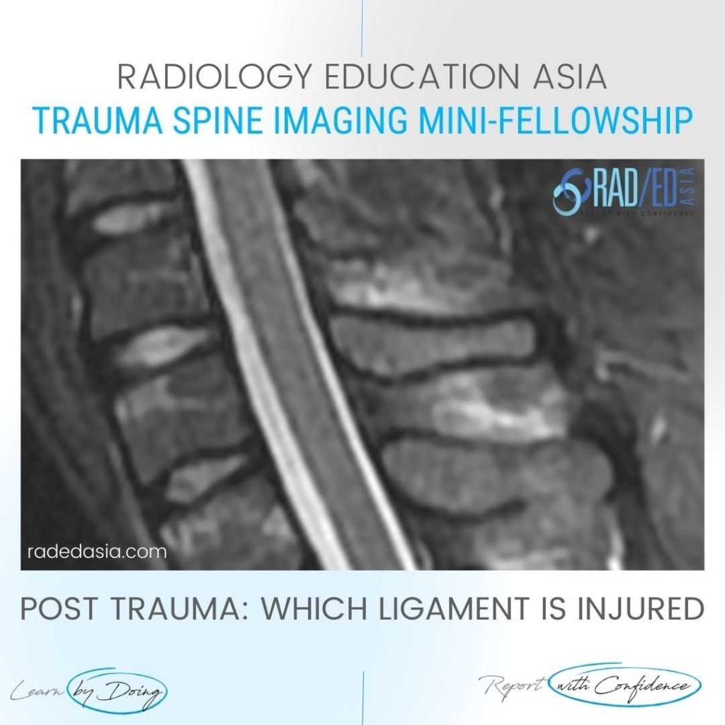
SPINE LIGAMENTS MRI INJURY RADIOLOGY TRAUMA INTERSPINOUS WHAT'S THE Dx: INTERSPINOUS LIGAMENT TRAUMA The MRI demonstrates trauma to the interspinous ligament with increased T2FS signal in the region of the interspinous ligament.   The interspinous ligament lies between the spinous processes.   Oedema indicates trauma to the region but is also a clue to the …

SPINE LIGAMENTS MRI INJURY RADIOLOGY TRAUMA INTERSPINOUS Read More »

vertebral artery dissection radiology mri diagnosis

VERTEBRAL ARTERY DISSECTION RADIOLOGY MRI DIAGNOSIS

VERTEBRAL ARTERY DISSECTION RADIOLOGY MRI DIAGNOSIS MRI VERTEBRAL ARTERY OCCULSION: DISCUSSION VERTEBRAL ARTERY OCCULSION   With spinal trauma its essential to look at the signal of the vertebral arteries as there can be dissection.   In most cases we don’t do a routine MRA so being able to spot a dissection on the standard scans …

VERTEBRAL ARTERY DISSECTION RADIOLOGY MRI DIAGNOSIS Read More »

chondromalacia patella femoral trochlear knee cartilage mri radiology radedasia

CHONDROMALACIA PATELLA FEMORAL TROCHLEAR CARTILAGE MRI KNEE

CHONDROMALACIA PATELLA FEMORAL TROCHLEAR CARTILAGE MRI DISCUSSION ON TROCHLEAR CARTILAGE MRI Chondromalacia of the femoral trochlear surface.   There is severe (in areas full thickness) cartilage loss (Green arrows) in the femoral trochlear.   Compare with normal thickness cartilage above and below (Pink arrows).   The femoral trochlear surface cartilage should be assessed on axial …

CHONDROMALACIA PATELLA FEMORAL TROCHLEAR CARTILAGE MRI KNEE Read More »

biceps tenosynovitis mri tendon sheath effusion tendinitis tendonitis radedasia

BICEPS TENDON SHEATH TENOSYNOVITIS EFFUSION MRI

BICEPS TENDON SHEATH TENOSYNOVITIS EFFUSION MRI DISCUSSION ON BICEPS TENOSYNOVITIS & TENDON SHEATH EFFUSION BICEPS TENDON SHEATH TENOSYNOVITIS   There can normally be a small amount of fluid in the biceps tendon sheath.   However in this case there is markedly increased fluid in the biceps tendon sheath in keeping with tenosynovitis.   Additionally there …

BICEPS TENDON SHEATH TENOSYNOVITIS EFFUSION MRI Read More »

bifurcate ligament radiology anterior mri anatomy

BIFURCATE LIGAMENT RADIOLOGY MRI ANATOMY ANKLE

BIFURCATE LIGAMENT RADIOLOGY MRI ANATOMY ANKLE DISCUSSION ON BIFURCATE LIGAMENT This is the Bifurcate ligament.   It arises from the anterior process of the calcaneum (Yellow arrow).   Its has two components that attaches: To the Navicular (Pink arrow). To the Cuboid (Green Arrow). KEY POINT ON BIFURCATE LIGAMENT On MRI, the Bifurcate Ligament is …

BIFURCATE LIGAMENT RADIOLOGY MRI ANATOMY ANKLE Read More »

This site is intended for Medical Professionals only. Use of this site is governed by our Terms of Service and Privacy Statement which can be found by clicking on the links. Please accept before proceeding to the website.
error: Right Click not available.