SEO

acromial spur mri shoulder radiology education asia

ACROMION / ACROMIAL SPURS ON MRI: WHERE TO LOOK AND WHAT TO LOOK FOR

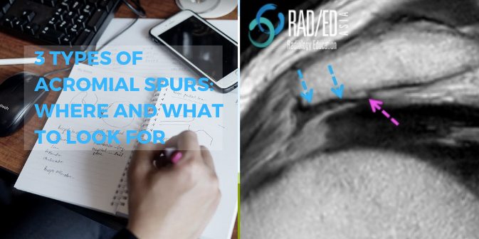
ACROMION SPURS ON MRI: 3 APPEARANCES ON MRI Acromial / Acromion spurs can be quite small and difficult to visualize on MRI. They are enthesophytes that arise from traction at the attachment sites of the Coraco Acromial Ligament and Deltoid. This post looks at the three locations and most common appearances of spurs arising from …

ACROMION / ACROMIAL SPURS ON MRI: WHERE TO LOOK AND WHAT TO LOOK FOR Read More »

freiberg disease mri radiology toe radedasia

FREIBERG DISEASE SUBCHONDRAL FRACTURE INFRACTION OSTEOCHONDROSIS (VIDEO)

FREIBERG DISEASE / SUBCHONDRAL FRACTURE INFRACTION / OSTEOCHONDROSIS DISCUSSION FREIBERG DISEASE / SUBCHONDRAL FRACTURE INFRACTION / OSTEOCHONDROSIS Extensive oedema in the head of the 2nd metatarsal with slight flattening of the cortical margin is characteristic of what’s called Freiberg’s Disease in adolescents and Subchondral Fracture in adults. Freiberg disease occurs in adolescents with an unfused …

FREIBERG DISEASE SUBCHONDRAL FRACTURE INFRACTION OSTEOCHONDROSIS (VIDEO) Read More »

mri plantar fascia tear fasciitis plantar fascia anatomy heel pain mri mri online course radedasia

MRI PLANTAR FASCIA ANATOMY: WHAT ARE THE 3 BANDS OF THE PLANTAR FASCIA (VIDEO)

MRI PLANTAR FASCIA ANATOMY MRI PLANTAR FASCIA ANATOMY: NORMAL BANDS Today’s post is a video on MRI of the Plantar Fascia Anatomy. We are looking at how to identify the 3 bands of the Plantar Fascia and which band is most important in Plantar Fasciitis. If you find the video helpful, please subscribe to the …

MRI PLANTAR FASCIA ANATOMY: WHAT ARE THE 3 BANDS OF THE PLANTAR FASCIA (VIDEO) Read More »

peroneal tubercle mri retrotrochlear eminence mri peroneal temdon tondonitis tear radiology education asia(1)

MSK MRI PERONEAL TUBERCLE & RETROTROCHLEAR EMINENCE MRI (VIDEO)

MRI PERONEAL TUBERCLE RETROTROCHLEAR EMINENCE MRI MSK How to identify the Peroneal tubercle and Retrotrochlear eminence on MRI: Today we are looking at MRI of the Peroneal Tubercle and the Retrotrochlear eminence. The importance of these structures is that when enlarged, a Peroneal Tubercle and less commonly the Retrotrochlear eminence, can result in Peroneal Tendonitis, …

MSK MRI PERONEAL TUBERCLE & RETROTROCHLEAR EMINENCE MRI (VIDEO) Read More »

acl tear mri segond fracture radiology education asia online radiology mri course

SEGOND FRACTURE MRI: WHERE TO LOOK, WHAT TO LOOK FOR, HOW TO REPORT (VIDEO)

MRI SEGOND FRACTURE KNEE ACL TEAR Segond Fracture MRI , How to find it when Assessing the ACL on MRI & what ACL associated injuries to look for: Today’s video post is on MRI of Segond Fracture which is one of the injuries associated with ACL tears. We look at where to look, what to …

SEGOND FRACTURE MRI: WHERE TO LOOK, WHAT TO LOOK FOR, HOW TO REPORT (VIDEO) Read More »

elbow mri brachialis tendon tears tendinosis radiology education asia

MRI ELBOW: MRI BRACHIALIS TENDON TEARS, TENDONOSIS AND NORMAL

MRI BRACHIALIS TENDON BRACHIALIS TENDON MRI: Elbow MRI Brachialis tendon Tendinosis, Tears and Normal   The Brachialis tendon is less commonly injured than the biceps.  It inserts onto the anterior ulnar on the ulnar tuberosity and to a lesser extent on the coronoid process but the tendon is very short compared to the biceps tendon. …

MRI ELBOW: MRI BRACHIALIS TENDON TEARS, TENDONOSIS AND NORMAL Read More »

acromial apophysiolysis apophysitis os acromiale radiology mri shoulder

ACROMIAL APOPHYSIOLYSIS OR APOPHYSITIS MRI FINDINGS

ACROMIAL APOPHYSIOLYSIS OR APOPHYSITIS MRI FINDINGS WHAT IS ACROMIAL APOPHYSIOLYSIS Acromial apophysiolysis or acromial apophysitis is a Stress injury due to repetitive traction injury at the acromial apophysis.   It results in instability at the apophysis and shoulder pain.   People who have acromial apophysitis have a higher incidence of eventually developing an Os Acromiale. …

ACROMIAL APOPHYSIOLYSIS OR APOPHYSITIS MRI FINDINGS Read More »

ankylosing spondylitis spine radiology ct mri fusion images

ANKYLOSING SPONDYLITIS SPINE RADIOLOGY CT MRI FUSION IMAGES

ANKYLOSING SPONDYLITIS SPINE RADIOLOGY CT MRI FUSION WHERE TO LOOK There are 4 sites in the posterior spine to assess for changes of AS. Facet Joints.  Superspinous Ligament.  Costovertebral joints.  Costo transverse joints. Changes can be at 1 or more of these sites. THE TAKE AWAY Spinal changes start anteriorly and progress posteriorly.   All …

ANKYLOSING SPONDYLITIS SPINE RADIOLOGY CT MRI FUSION IMAGES Read More »

This site is intended for Medical Professionals only. Use of this site is governed by our Terms of Service and Privacy Statement which can be found by clicking on the links. Please accept before proceeding to the website.
error: Right Click not available.