

knee mri deep mcl meniscotibial ligament tear dmcl radiology

MENISCOTIBIAL LIGAMENT TEAR DEEP MCL MRI KNEE

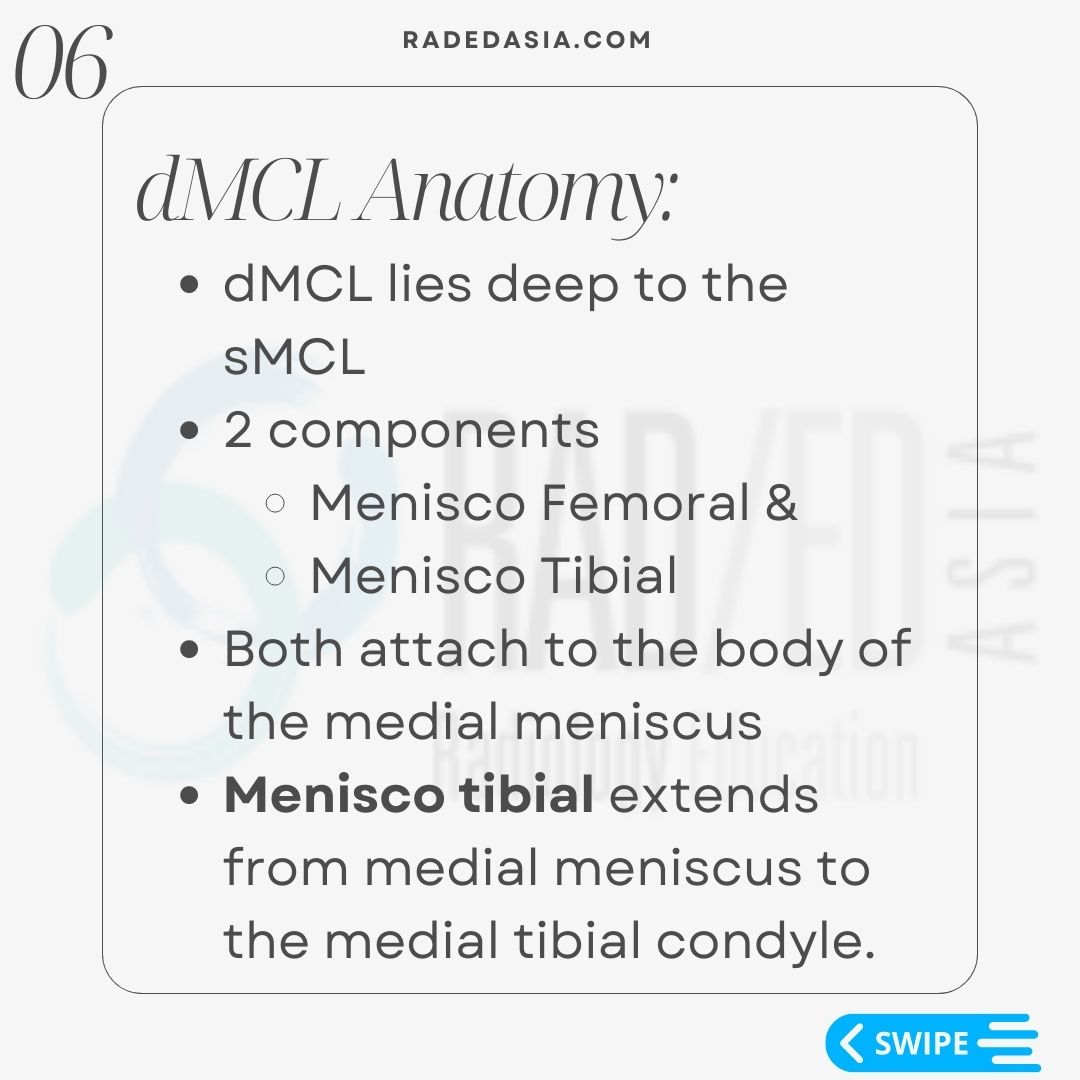
  • The dMCL lies deep to the sMCL.
  • It has 2 components.
    1. Menisco Femoral &,
    2. Menisco Tibial.
  • Both attach to the body of the medial meniscus
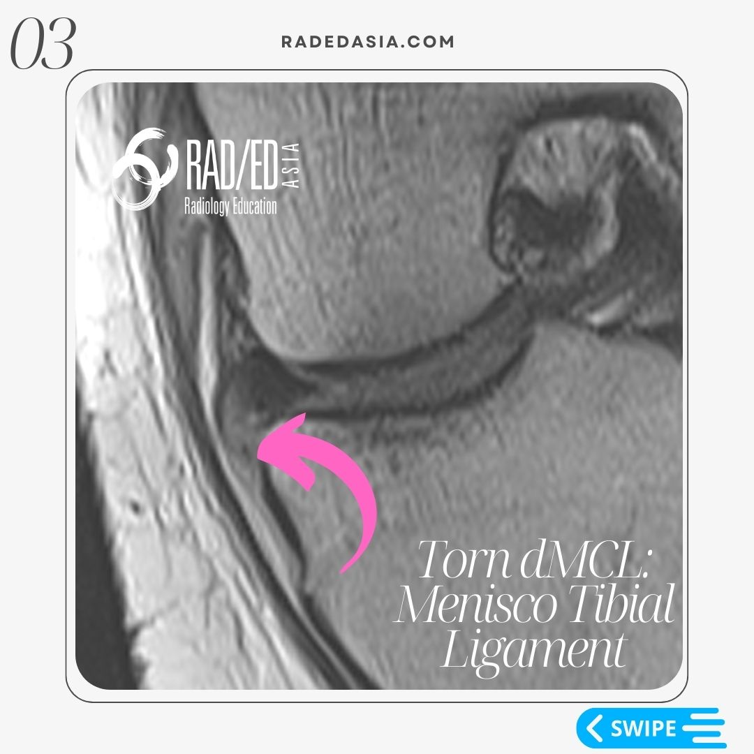
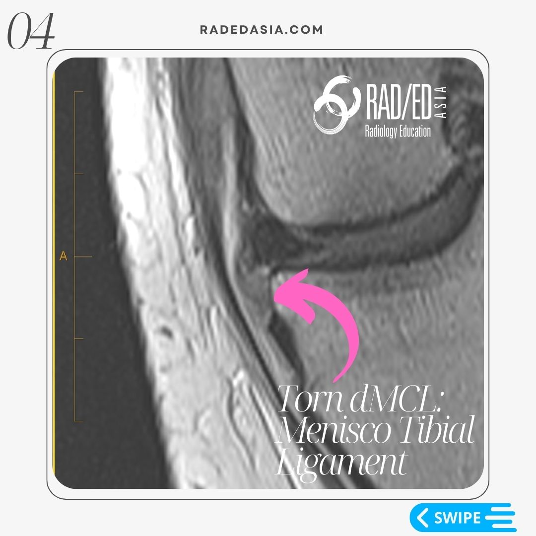
  • The Meniscotibial ligament extends from medial meniscus to the medial tibial condyle.

  • Look for 2 things,
    1. Increased signal of the ligament.
      • Normally the menisco-tibial ligament is low signal.
    2. Ill definition & thickening of the ligament.
  • The menisco-tibial component is stronger than the menisco-femoral and is less commonly torn.

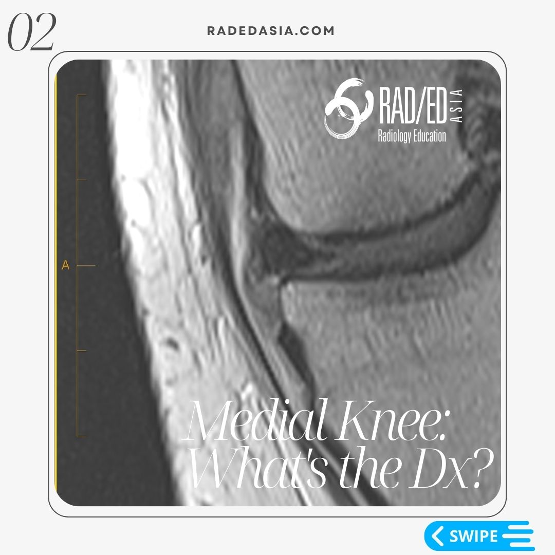
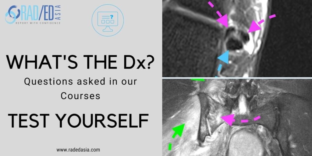
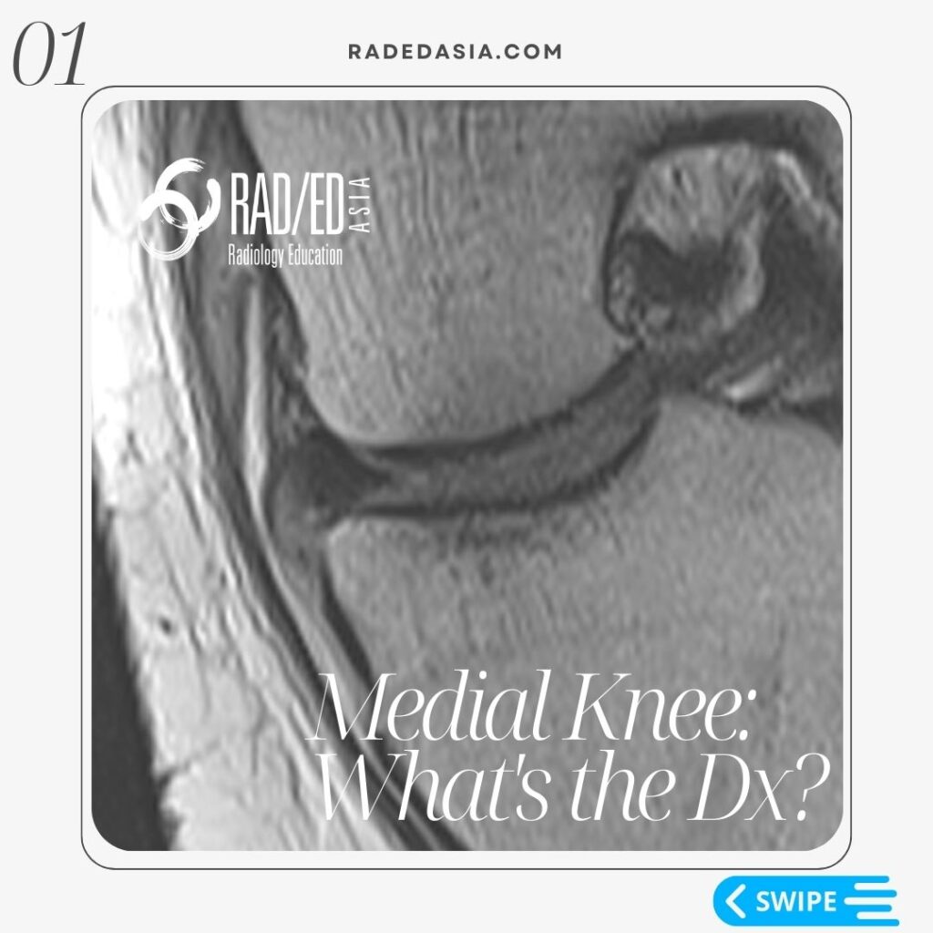
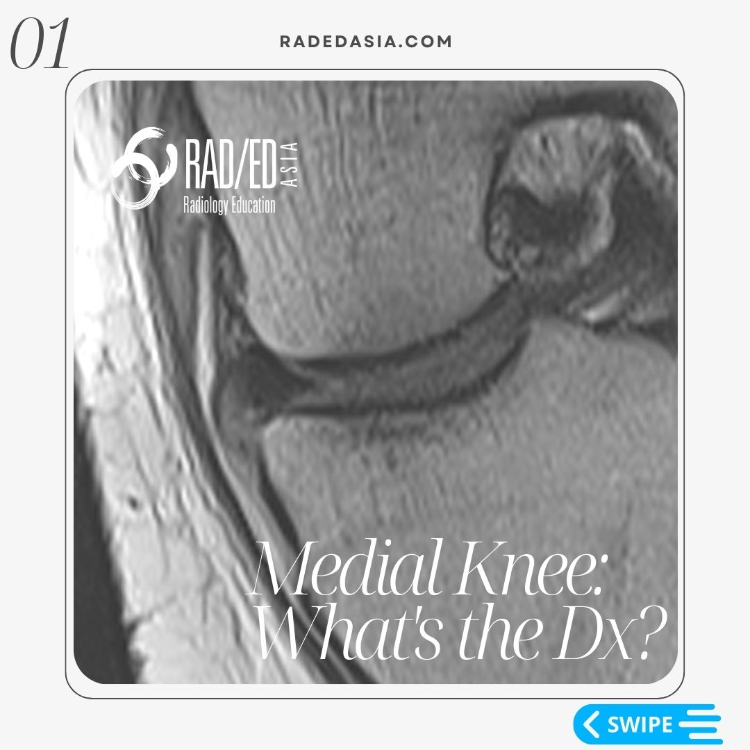
In this case the menisco-tibial ligament is hyper-intense, thickened and ill defined (Pink arrows). All features of a torn menisco-tibial ligament.

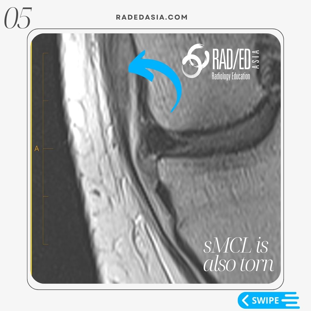
You can also see that similar changes are present in the proximal sMCL which is also torn (Blue arrow).
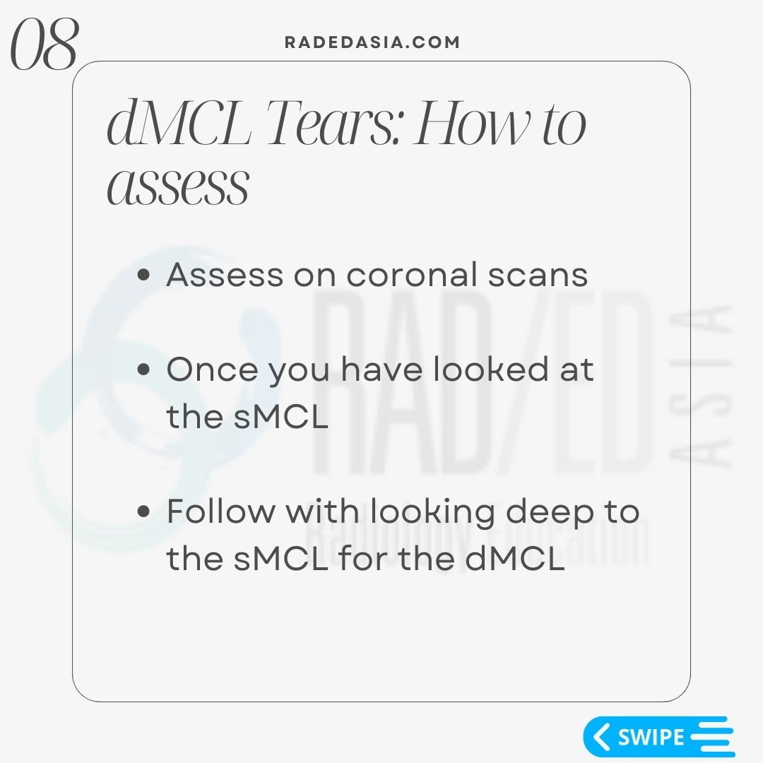
  • Assess on coronal scans the menisco-tibial and menisco-femoral ligaments.
     
  • Start by assessing the sMCL.
     
  • Once you have looked at the sMCL look immediately deep to it to find the deep MCL components.

Learn more about Knee Imaging in our ONLINE
Guided MRI KNEE Mini-Fellowship.


More by clicking on the images below

Estructuras de la rodilla afectadas por la CPPD de la rodilla, incluyendo fibrocartílago, cartílago, tendones y ligamentos.
INFORMES
Fracturas laterales de la rodilla y las estructuras ligamentarias y tendinosas involucradas en la RM de rodilla.
FRACTURA
error: Content is protected !!

Suscríbase a nuestro boletín

Esté atento a los nuevos lanzamientos y aprendizajes de las Fellowships

Fellowships

Mejor que las conferencias

Únase a nuestra comunidad de WhatsApp

Ayudamos a los radiólogos a aprender
haciendo e informa con confianza

Este sitio está destinado exclusivamente a las profesiones médicas. El uso de este sitio se rige por nuestras Condiciones de servicio y Declaración de privacidad, que puede consultar haciendo clic en los enlaces. Por favor, acéptelas antes de continuar en el sitio web.