knee mri deep mcl meniscotibial ligament tear dmcl radiology
  • The dMCL lies deep to the sMCL.
     
  • It has 2 components.
    1. Menisco Femoral &,
    2. Menisco Tibial.


  • Both attach to the body of the medial meniscus


  • The Meniscotibial ligament extends from medial meniscus to the medial tibial condyle.

  • Look for 2 things,
    1. Increased signal of the ligament.
      • Normally the menisco-tibial ligament is low signal.
    2. Ill definition & thickening of the ligament.


  • The menisco-tibial component is stronger than the menisco-femoral and is less commonly torn.

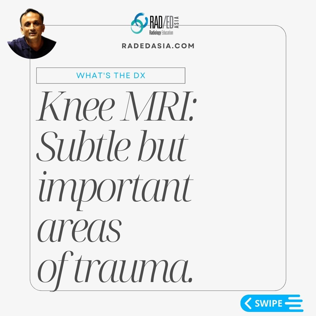
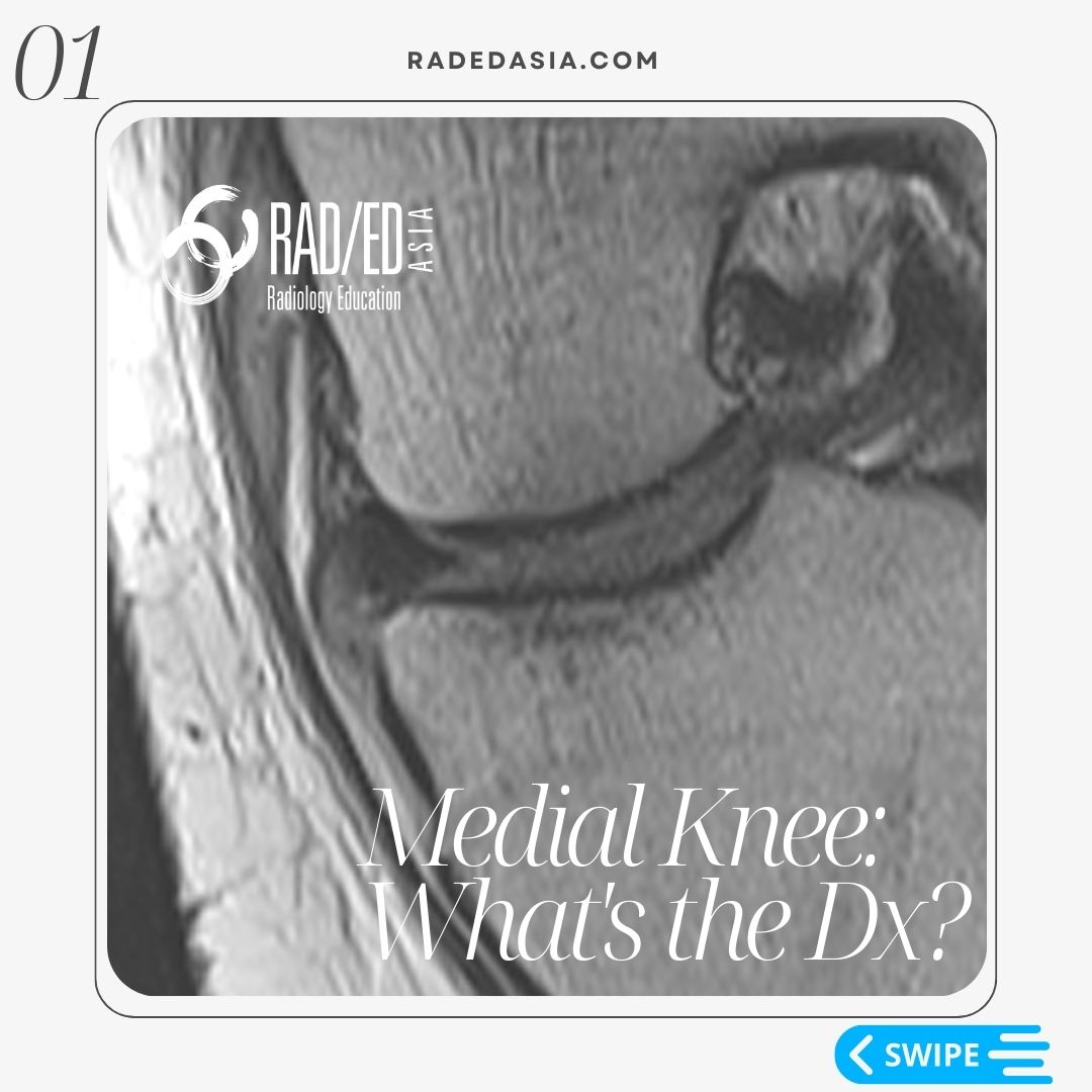
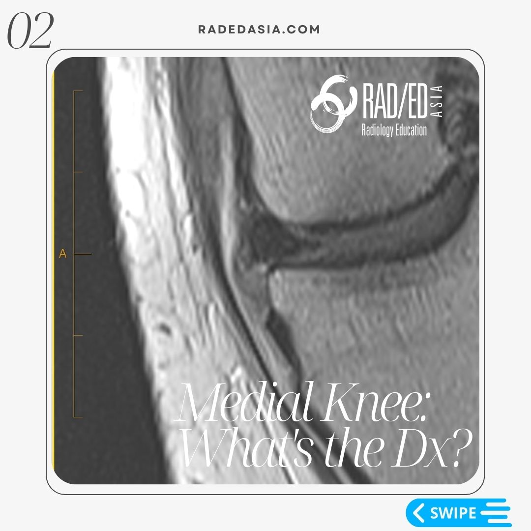
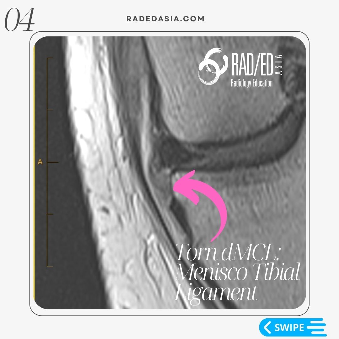
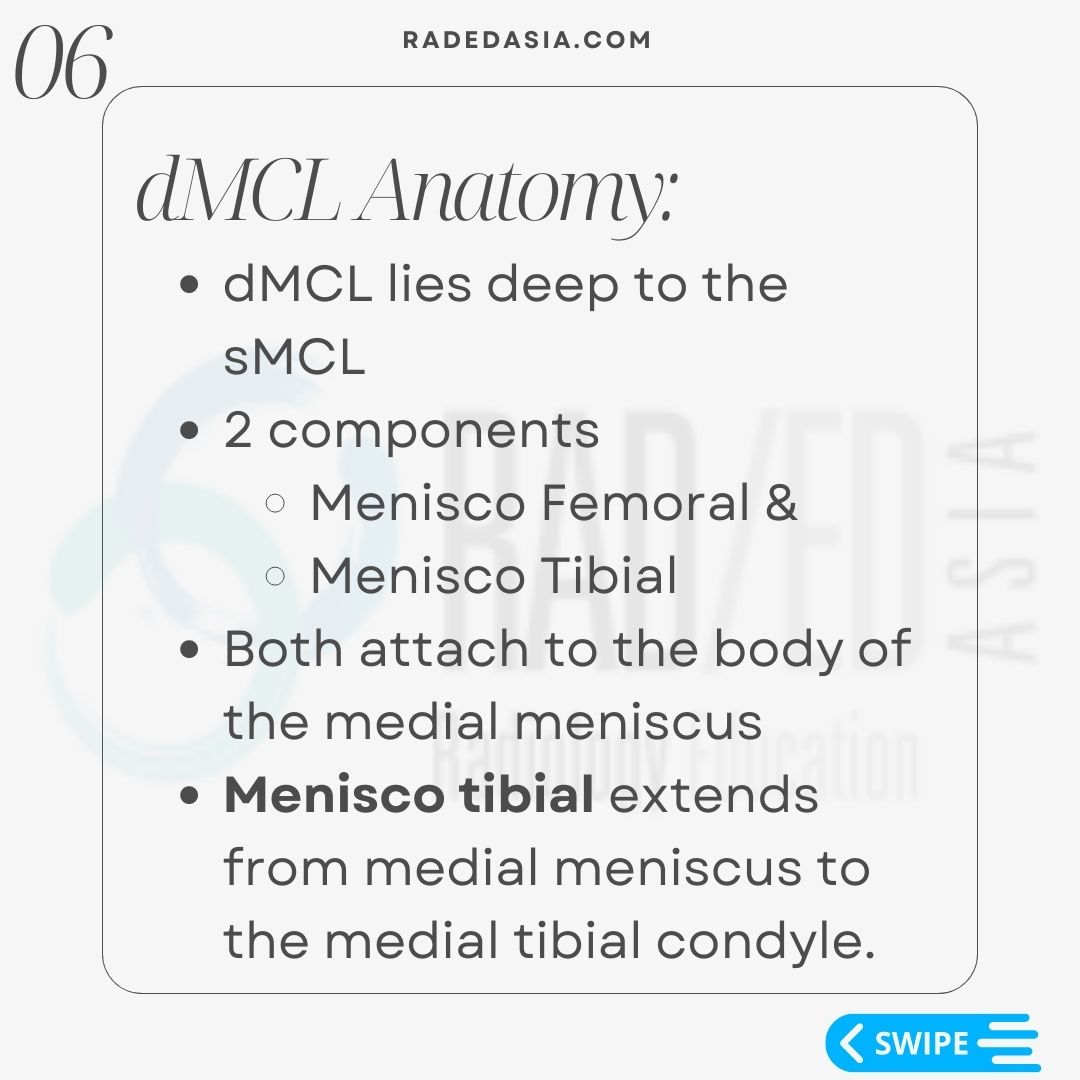
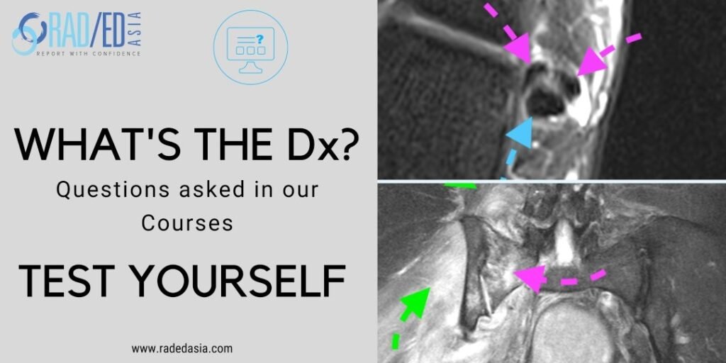
In this case the menisco-tibial ligament is hyper-intense, thickened and ill defined (Pink arrows). All features of a torn menisco-tibial ligament.


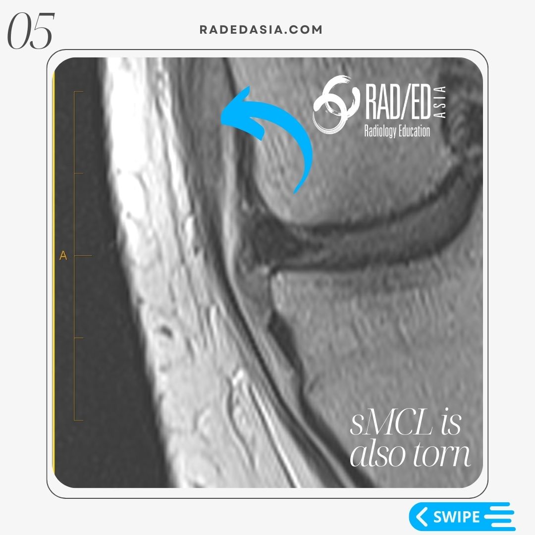
You can also see that similar changes are present in the proximal sMCL which is also torn (Blue arrow).

  • Assess on coronal scans the menisco-tibial and menisco-femoral ligaments.
     
  • Start by assessing the sMCL.
     
  • Once you have looked at the sMCL look immediately deep to it to find the deep MCL components.

Learn more about KNEE Imaging in our ONLINE or ONSITE
Guided MRI KNEE Mini-Fellowship.


More by clicking on the images below.

acl-pcl-mucoid-degeneration-mri-findings-radedasia
Anterior Cruciate Ligament
mri knee meniscus degeneration maceration flap tear extrusion meniscocapsular separation radedasia
KNEE

#radiology #radedasia #mri #kneemri #msk #mriknee #mskmri #radiologyeducation #radiologycases #radiologist #rads #radiologystudent #radiologycme #radiologycpd #medicalimaging #imaging #radcme #rheumatology #arthritis #rheumatologist #sportsmed #orthopaedic #physio #physiotherapist #MCL #dMCL #sMCL

#radedasia #mri #mskmri #radiology

This site is intended for Medical Professionals only. Use of this site is governed by our Terms of Service and Privacy Statement which can be found by clicking on the links. Please accept before proceeding to the website.
error: Right Click not available.