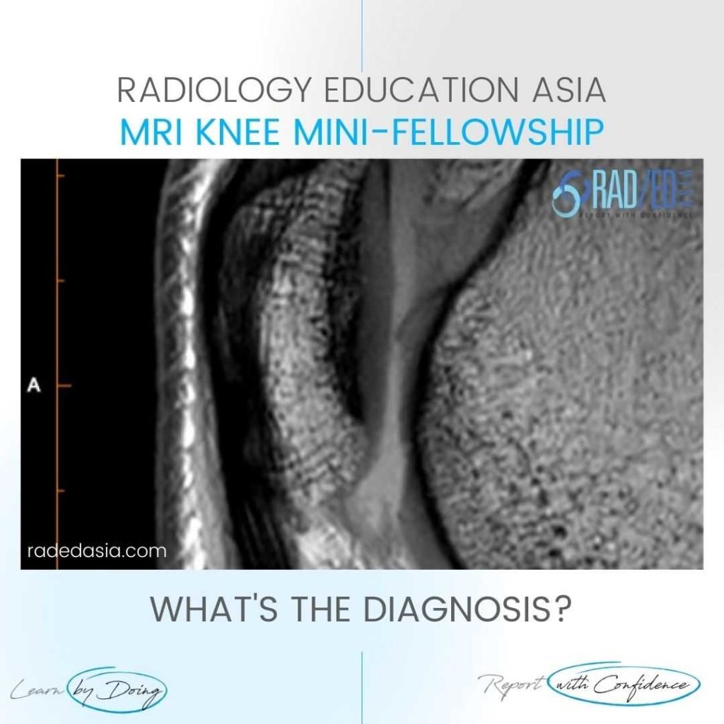
VAUGHN JACKSON SYNDROME WRIST MRI
Vaughn Jackson Syndrome in Rheumatoid Arthritis: Wrist MRI Online Radiology Course This case is from Dr Joe Thomas a senior consultant Rheumatologist who is also involved with our Spine Arthropathy and Spondyloarthropathy course and our upcoming Peripheral Arthropathy Course. Thanks to Dr. Binoy P S Specialist Hand Reconstructive Surgery and Microsurgery CLINICO RADIOLOGICAL CORRELATION CLINICAL …