neuropathic joint-syrinx syringomyelia spine mri online radiology courses

NEUROPATHIC JOINT ELBOW SYRINX SYRINGOMYELIA SPINE MRI

radiology rheumatology imaging dr joe thomas

A combined Clinico Radiological case from Dr Joe Thomas (Rheumatologist)

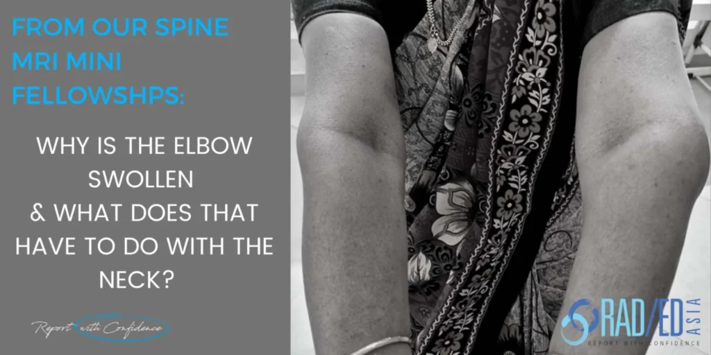
Neuropathic Elbow Joint due to a Syrinx. A case from Dr Joe Thomas a senior consultant Rheumatologist who is also joining us in presenting the Spine Arthropathy and Spondyloarthropathy course.

neuropathic joint-syrinx syringomyelia spine mri radedasia

  • Right handed 60 yo lady presented with left elbow swelling and mild pain. No movement restriction.
  • No other joints involved.
  • Negative for inflammatory arthropathy workup.

neuropathic joint-syrinx syringomyelia xray left elbow spine mri radedasia

Image above: Multiple loose bodies and effusion. No joint destruction. No chondrocalcinosis.

neuropathic joint-syrinx syringomyelia mri left elbow spine mri radedasia

Image above: Effusion(Pink arrow) Extensive full thickness cartilage loss (Green arrows) Subcortical geode (Orange arrow) and Extensive generalized bone marrow oedema (Blue arrows).

RADIOLOGICAL INTERPRETATION:

The imaging findings are those of a severe degenerative changes with multiple loose bodies. An unusual finding for OA was the extensive generalised bone marrow oedema.

WHAT WAS THE CLINICAL INTERPRETATION OF THE IMAGING FINDINGS

1. Imaging suggests extensive degeneration but the elbow is an uncommon joint for primary OA. Additionally the patient is right handed and this is the left elbow involved.

2. Based on degenerative appearing changes in the elbow, an unusual joint for primary OA, the main clinical differential considered by Dr Thomas was either Neuropathic arthropathy or CPPD.

3. CPPD was thought unlikely bases on the lack of a typical appearance and absence of any other areas of involvement. 

So a Cervical MRI was performed.

neuropathic joint-syrinx syringomyelia mri cord radedasia

Image above: Benign syrinx cervical cord (Pink arrows) larger to left side.

The patient was also seen by a Neurologist and based on the combination of Imaging and Clinical findings, a Neuropathic arthropathy due to a syrinx was diagnosed.

  • The classical appearance of a neuropathic joint is of bone destruction, disorganisation and loose bodies and in this case only the loose bodies are present.

  • There is however an earlier phase of neuropathic arthropathy where the appearance is of joint degeneration without bone destruction.

  • There are many causes of Neuropathic arthropathy.
  • But in the shoulder and elbow, the most common cause of a neuropathic arthropathy is a syrinx.

There are two theories on how changes occur in a neuropathic joint:

    • Repetitive trauma to a joint that has no sensation leads to joint destruction.

    • Dysfunction of the sympathetic system results in excess bone resorption resulting in pathological fractures.


Neuropathic joint can develop either early or late in the syrinx formation.

  • Two forms of Neuropathy Arthropathy are described
  • ATROPHIC: Extensive bone resorption at the joint.
  • HYPERTROPHIC: Joint destruction with new bone formation, loose bodies, sclerosis, fractures.

  • Upper limb neuropathic arthropathy usually caused by a syrinx.

  • Most commonly shoulder or elbows.

  • Uncommon for wrist or multiple joint involvement.

  1. An early appearance of neuropathic arthropathy may be severe degenerative type change.

  2. Seeing this in the elbow should raise the suspicion of a neuropathic arthropathy.

  3. If there is evidence or suspicion of a neuropathic joint in the upper limb make sure the cord is assessed on MRI for a syrinx.

Learn more about this condition & How best to report it in more detail in our SPINE MRI Mini Fellowships.

Click on the image below for more information.

Our upcoming onsite Guided MSK MRI Mini Fellowships.

Click on the image below for more information.

For all our other current MSK MRI & Spine MRI
Online Guided Mini Fellowships.

Click on the image below for more information.

baastrup-disease-ct-findings-radedasia
PAIN MANAGEMENT
spine-imaging-course-pain-management-sports-medicine-orthopaedic-specialists-ct-mri
ONLINE MINI-FELLOWSHIPS

#radiology #radiologist #radiologia #mri #spinermri #mskmri #msk #mskrad #mskradiology #imaging #frcr #sportsmed #radiologyresident #foamrad #ortho #ct #radiologystudent #trauma #radedasia #radiologycme #radiologyeducation #radiologycases #rheumatology #arthritis #painphysician #chiropractic #physiotherapy#spondyloarthritis #ankylosingspondylitis #spinearthritis #emergencydepartment #spinemri #cordmri #mrispine #charcots #neuropathic joint #rheumatology #rheumatologist

#radedasia #mri #mskmri #radiology

This site is intended for Medical Professionals only. Use of this site is governed by our Terms of Service and Privacy Statement which can be found by clicking on the links. Please accept before proceeding to the website.
error: Right Click not available.