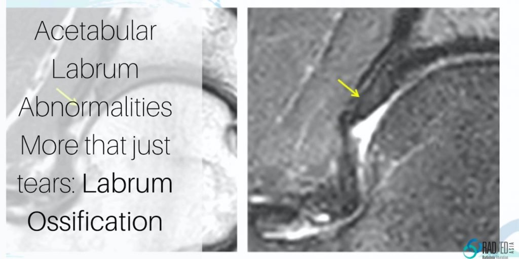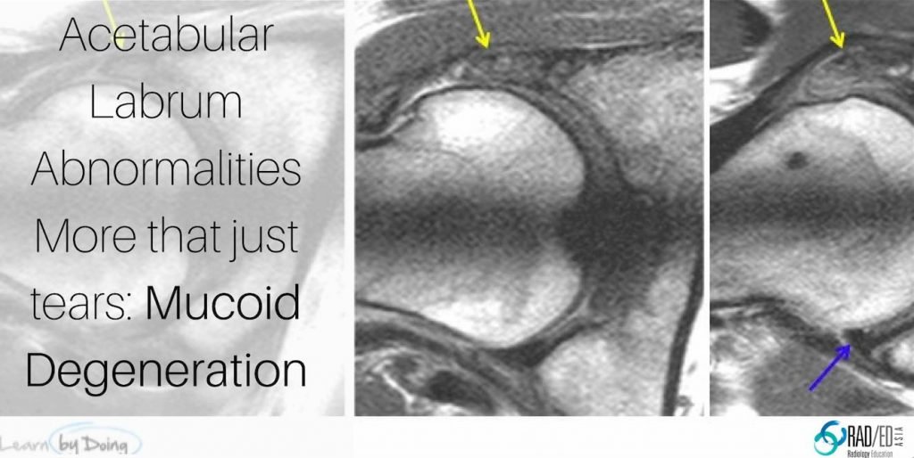
A FEW HIP LABRUM ABNORMALITIES ON MRI & CT
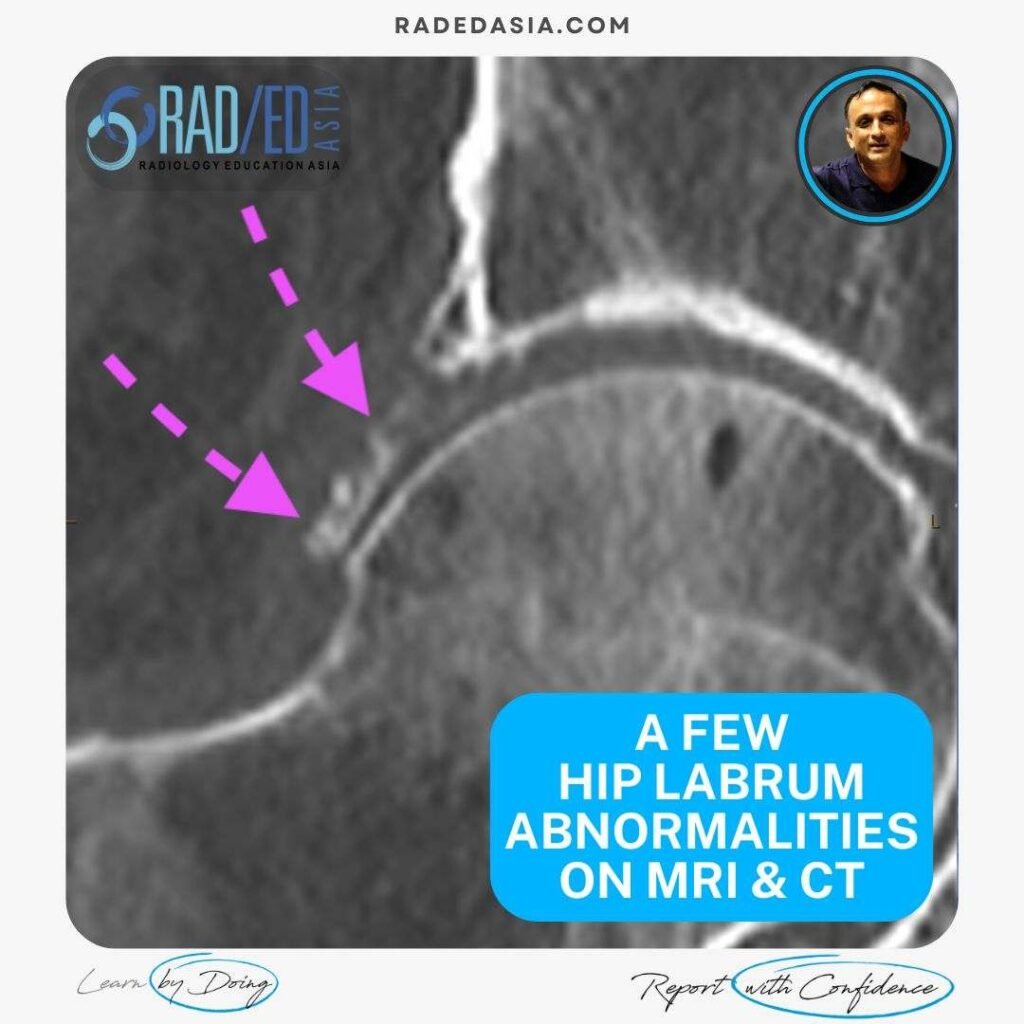
HIP LABRUM ABNORMALITIES ON MRI & CT HIP LABRUM MRI & CT There is more to the hip labrum than just Labral tears. Here is a Quick Look at a few Hip Labrum abnormalities from Labral tears, paralabral cysts and mucoid degeneration to Labral ossification. A FEW HIP LABRUM ABNORMALITIES LABRAL TEAR WITH PARALABRAL …