PAIN MANAGEMENT

Pain management Physician Anesthetist

All our posts on pain management are done in conjunction with Dr Rajendra Sahoo is a senior consultant Pain Physician and Anesthetist who is Fellowship trained in Ultrasound guided Pain Management & has a vast amount of clinical experience.

We will be collaborating with Dr Sahoo to bring more MSK and Spine Ultrasound learning.

mri-baastrup's-disease-imaging-findings-what-to-look-for-radedasia

MRI BAASTRUP’S DISEASE: WHAT TO LOOK FOR

BAASTRUP'S DISEASE: WHAT TO LOOK FOR ON MRI Baastrup’s disease is caused by the friction of two spinous processes rubbing against each other and can result in central back pain but its difficult to diagnose without imaging. In the previous post we looked at the CT Findings in Baastrup’s Disease and this post looks at …

MRI BAASTRUP’S DISEASE: WHAT TO LOOK FOR Read More »

acl-pcl-mucoid-degeneration-mri-findings-radedasia

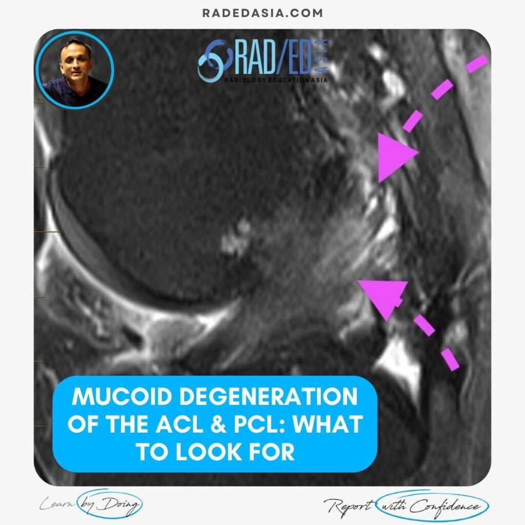
ACL & PCL MUCOID DEGENERATION MRI FINDINGS

MRI FINDINGS IN ACL AND PCL MUCOID DEGENERATION Mucoid degeneration can affect both the anterior cruciate ligament (ACL) and posterior cruciate ligament (PCL). Mucoid degeneration is often confused with ACL tears and this post looks at the main MRI features of ACL and PCL mucoid degeneration that will help to differentiate from tears. MRI FINDINGS …

ACL & PCL MUCOID DEGENERATION MRI FINDINGS Read More »

mri knee meniscus degeneration maceration flap tear extrusion meniscocapsular separation radedasia

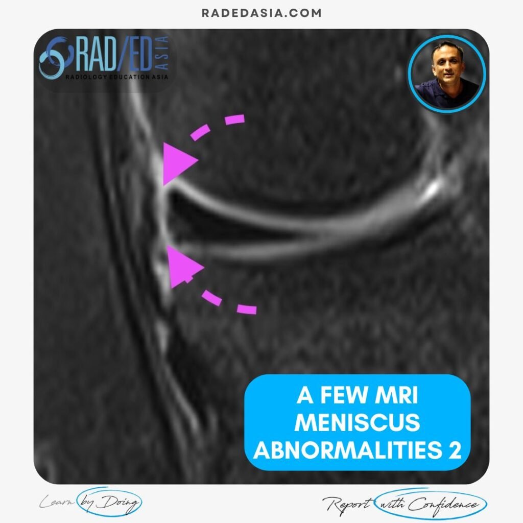
A FEW MRI MENISCUS ABNORMALITIES 2: MRI MENISCUS DEGENERATION MACERATION FLAP TEAR EXTRUSION AND MENISCOCAPSULAR SEPARATION

MENISCUS ABNORMALITIES 2 KNEE MRI MRI MENISCUS ABNORMALITIES A few more MRI Meniscus abnormalities such as meniscus maceration, flap tears, extrusion and meniscocapsular separation. MRI MENISCUS ABNORMALITIES MENISCO CAPSULAR SEPARATION MRI Meniscocapsular separation is a separation of the attachment of the external margin of the body of the medial meniscus from the Posterior Oblique ligament. …

A FEW MRI MENISCUS ABNORMALITIES 2: MRI MENISCUS DEGENERATION MACERATION FLAP TEAR EXTRUSION AND MENISCOCAPSULAR SEPARATION Read More »

spine-imaging-course-pain-management-sports-medicine-orthopaedic-specialists-ct-mri

SPINE & SIJ IMAGING FOR PAIN MANAGEMENT AUGUST 2024

Home SPINE & SIJ IMAGING COURSE FOR PAIN MANAGEMENT GUIDED MRI & CT SPINE IMAGING COURSE FOR PAIN MANAGEMENT: 10th AUGUST 2024 If you work in Spine Pain Management and have not had formal training in assessing Spine MRI and CT, our new GUIDED SPINE & SIJ IMAGING FOR PAIN MANAGEMENT Mini Fellowship will help …

SPINE & SIJ IMAGING FOR PAIN MANAGEMENT AUGUST 2024 Read More »

mri knee meniscus tears cysts pathologies images

A FEW MRI MENISCUS ABNORMALITIES: MENISCUS TEAR, INTRA MENISCAL CYST AND VARIANTS THAT LOOK LIKE TEARS

MENISCUS ABNORMALITIES KNEE MRI MRI MENISCUS ABNORMALITIES A few MRI Meniscus abnormalities such as Meniscus tear, Intra meniscal cyst and variants that look like tears. MRI MENISCUS ABNORMALITIES MRI MENISCUS INTRAMENISCAL CYST WHAT ARE THE FINDINGS? Cystic structure (Pink arrow) present within the anterior horn. WHAT’S THE Dx? INTRAMENISCAL CYST Extensive intra meniscal cyst anterior …

A FEW MRI MENISCUS ABNORMALITIES: MENISCUS TEAR, INTRA MENISCAL CYST AND VARIANTS THAT LOOK LIKE TEARS Read More »

mri achilles tendon abnormalities ankle radiology

MRI OF A FEW ACHILLES TENDON ABNORMALITIES: INSERTIONAL TENDONOPATHY TENDONOSIS, BURSA & RUPTURE

ACHILLES TENDON ABNORMALITIES ANKLE MRI ACHILLES TENDON ABNORMALITIES A few MRI images of Achilles pathology from all the findings seen in Achilles Insertional Tendonopathy, Retrocalcaneal Bursitis, Retroachilles Bursitis to MRI of Achilles Rupture. MRI ACHILLES TENDON ABNORMALITIES MRI ACHILLES INSERTIONAL TENDONOPATHY: WHAT TO LOOK FOR MRI findings in Insertional Achilles Tendinopathy: A few findings to …

MRI OF A FEW ACHILLES TENDON ABNORMALITIES: INSERTIONAL TENDONOPATHY TENDONOSIS, BURSA & RUPTURE Read More »

pyriformis syndrome pudendal nerve quadratus femoris ischiofemoral impingement ultrasound scan inject radedasia

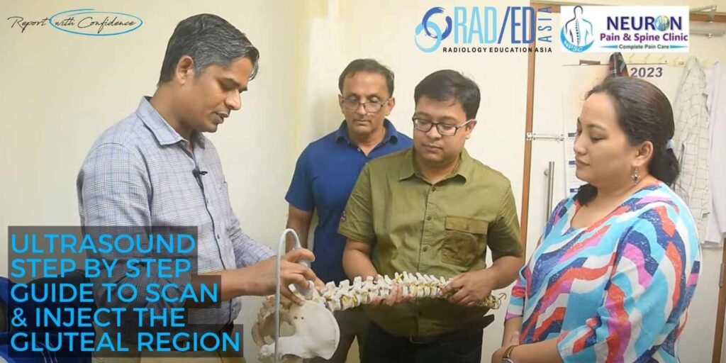
ULTRASOUND PAIN MANAGEMENT HOW TO SCAN AND INJECT FOR PIRIFORMIS SYNDROME, PUDENDAL NERVE, QUADRATUS FEMORIS & ISCHIOFEMORAL IMPINGEMENT

STEP BY STEP GUIDE ON HOW TO SCAN AND INJECT THE GLUTEAL REGION FOR PAIN MANAGEMENT HOW TO SCAN AND INJECT THE GLUTEAL REGION FOR PAIN MANAGEMENT   In this video by Dr Rajendra Sahoo Pain Management Specialist at KIMS Hospital and Neuron Pain Clinic we look at the role of ultrasound in the diagnosis …

ULTRASOUND PAIN MANAGEMENT HOW TO SCAN AND INJECT FOR PIRIFORMIS SYNDROME, PUDENDAL NERVE, QUADRATUS FEMORIS & ISCHIOFEMORAL IMPINGEMENT Read More »

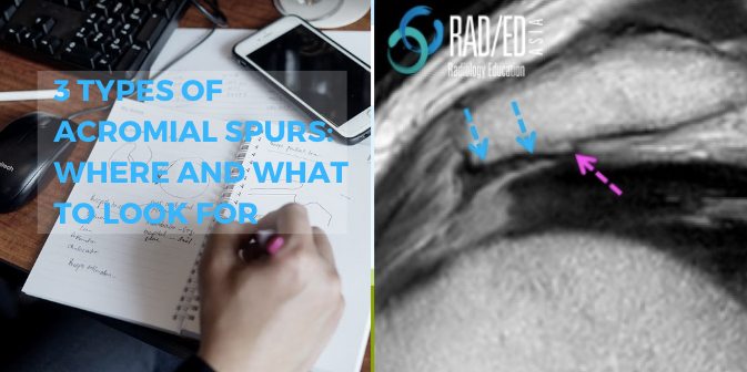
acromial spur mri shoulder radiology education asia

ACROMION / ACROMIAL SPURS ON MRI: WHERE TO LOOK AND WHAT TO LOOK FOR

ACROMION SPURS ON MRI: 3 APPEARANCES ON MRI Acromial / Acromion spurs can be quite small and difficult to visualize on MRI. They are enthesophytes that arise from traction at the attachment sites of the Coraco Acromial Ligament and Deltoid. This post looks at the three locations and most common appearances of spurs arising from …

ACROMION / ACROMIAL SPURS ON MRI: WHERE TO LOOK AND WHAT TO LOOK FOR Read More »

This site is intended for Medical Professionals only. Use of this site is governed by our Terms of Service and Privacy Statement which can be found by clicking on the links. Please accept before proceeding to the website.
error: Right Click not available.