A FEW MRI MENISCUS ABNORMALITIES 2: MRI MENISCUS DEGENERATION MACERATION FLAP TEAR EXTRUSION AND MENISCOCAPSULAR SEPARATION
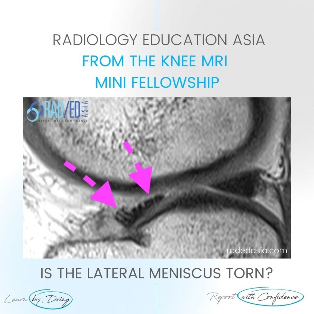
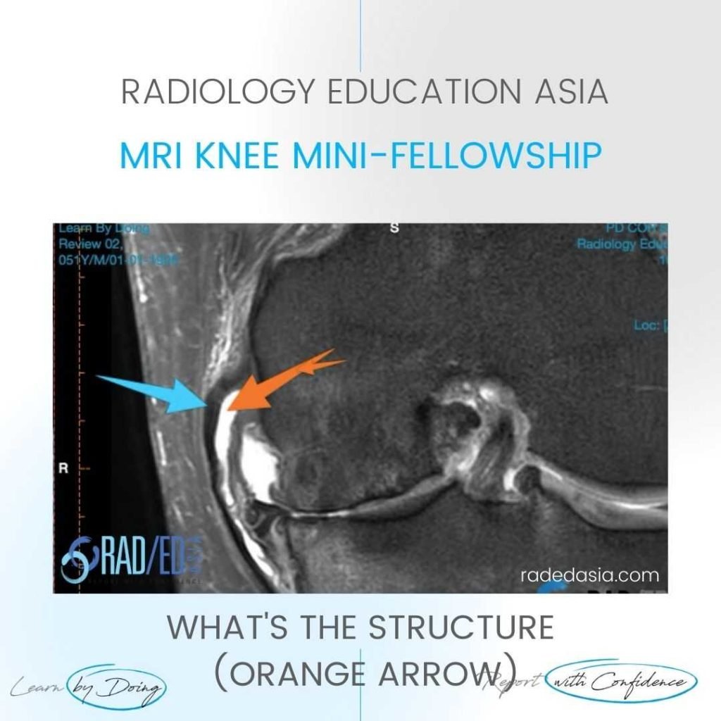
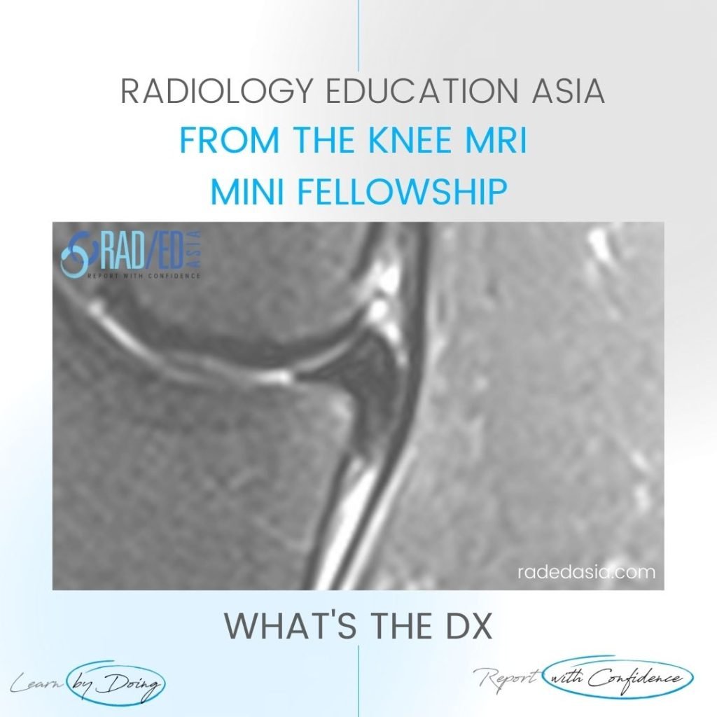
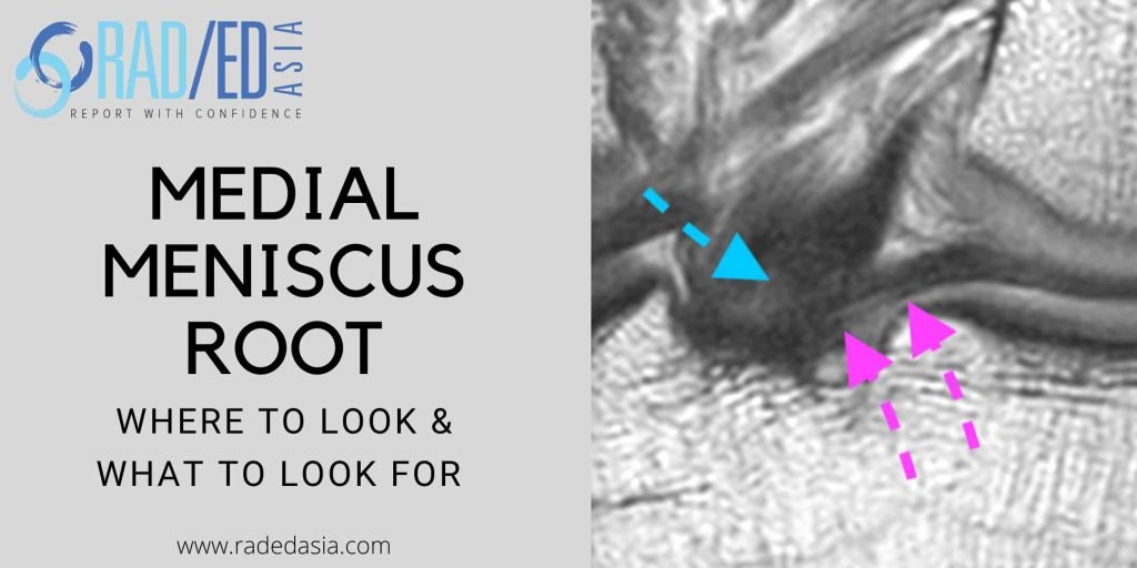
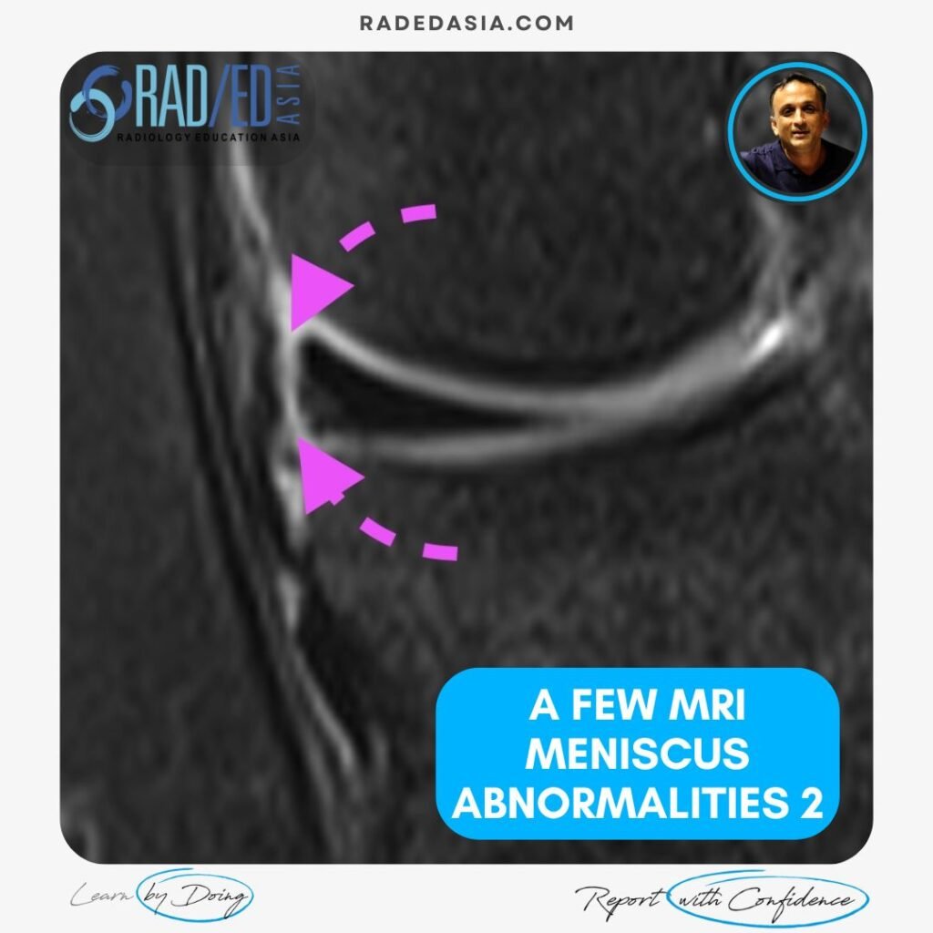
MENISCUS ABNORMALITIES 2 KNEE MRI MRI MENISCUS ABNORMALITIES A few more MRI Meniscus abnormalities such as meniscus maceration, flap tears, extrusion and meniscocapsular separation. MRI MENISCUS ABNORMALITIES MENISCO CAPSULAR SEPARATION MRI Meniscocapsular separation is a separation of the attachment of the external margin of the body of the medial meniscus from the Posterior Oblique ligament. …