This site is intended for Medical Professions only. Use of this site is governed by our Terms of Service and Privacy Statement which can be found by clicking on the links. Please accept before proceeding to the website.

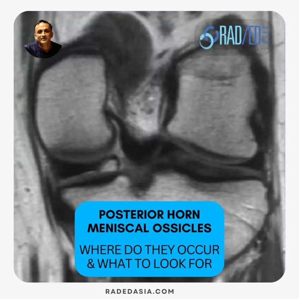
Meniscal ossicles are areas of ossification that occur within the meniscus.


They can occur anywhere in either meniscus but the most common location for a meniscal ossicle to occur is in the meniscal root or posterior horn / root junction of the medial meniscus.
They can also be seen in the lateral meniscus but this is very uncommon.
Here is an abridged version of the location they are seen in from Mohankumar et al’s article (Link in previous tab):

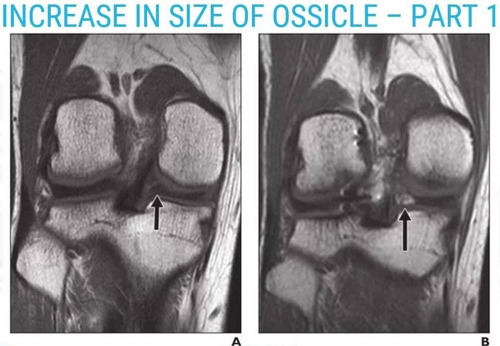
Meniscal ossicles can increase in size on sequential scans.
The two images below (From Mohankumar et al’s previously credited article) demonstrate.
1. Interval development of ossicle.
Image Above: Image A initial scan normal posterior horn/root. Image B 33 months later development of meniscal ossicle (arrow). Images from credited article.
2. Interval increases in size.

Image Above: Temporal increase in size of ossicle. Initial scan (Image A) shows small ossicle (Arrow) which 18 months later (Image B) has increased in size. Images from credited article.

Because ossicles follow fat signal when you fat saturate, they will appear black and will have the same signal as adjacent meniscus and you are likely to miss them.
Always look also at the non-fat saturated scans.

Meniscal ossicles are areas of ossification that occur within the meniscus.

Meniscal ossicles can occur either post-traumatically as a result of metaplasia and heterotopic ossification of a meniscal tear, or due to avulsion of bone related to a meniscal root tear.

The most common location for meniscal ossicles is in the meniscal root or posterior horn of the medial meniscus.
Meniscal ossicles appear iso-intense to bone marrow on all sequences of imaging.


Stay tuned on new
Mini-Fellowships launches and learnings
This site is intended for Medical Professions only. Use of this site is governed by our Terms of Service and Privacy Statement which can be found by clicking on the links. Please accept before proceeding to the website.