Spondyloarthropathy
MRI spondyloarthropathy spine
Imaging of Ankylosing Spondylitis and Spondyloarthritis can be challenging because the clinical context of what we are seeing is very important in getting to or narrowing a diagnosis.

From 2021 all our posts on Imaging and MRI Ankylosing Spondylitis and Spondyloarthropathies will be done in conjunction with Dr Joe Thomas a Senior Consultant Rheumatologist based in Kochi India, who has a vast amount of clinical experience.
He also has a very strong interest in Imaging of Spondyloarthritis and has joined us to bring a clinical perspective to the imaging and to advise on what rheumatologists want when we report their referrals.
MRI Spondyloarthropathy and Ankylosing Spondylitis. Where to Look, What to Look for and How to Report Confidently
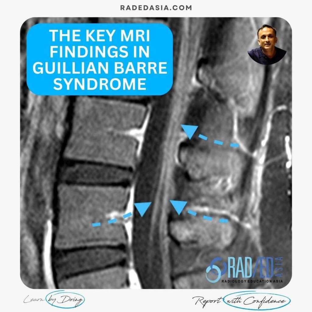
WHAT ARE THE MRI FINDINGS IN GUILLIAN- BARRE SYNDROME Guillain-Barré Syndrome (GBS) has distinct MRI findings in the spine. In this post we look at the characteristic MRI findings of GBS in the spine. WHAT ARE KEY MRI FINDINGS TO LOOK FOR IN GUILLAIN-BARRÉ SYNDROME (GBS)? NERVE ROOT ENHANCEMENT This is the most significant MRI …
MRI FINDINGS IN GUILLIAN BARRE SYNDROME Read More »
BASILAR INVAGINATION MRI RHEUMATOID ARTHRITIS BASILAR INVAGINATION MRI RHEUMATOID ARTHRITIS MCRAE'S LINE There can be apparent superior migration of the dens to the foramen magnum. It’s apparent because it’s due to cranial settling with the skull base moving down rather than the odontoid moving up. To assess this on imaging, there are a …
BASILAR INVAGINATION MRI RHEUMATOID ARTHRITIS MCRAE’S LINE (VIDEO) Read More »
ROMANUS LESIONS ANATOMY OF THE ENDPLATE AND ENTHESIS To understand the Romanus Lesion or Corner Inflammatory Lesion as its described on MRI, its important to understand the anatomy of the endplate and enthesis it forms with the annulus. This post was done together with Dr Joe Thomas who is a very talented Rheumatologist from Kochi …
ROMANUS LESIONS ANATOMY OF THE ENDPLATE AND ENTHESIS Read More »
ROMANUS LESION X-RAY CT FINDINGS Romanus Lesion is a term used on x-ray and CT and on MRI is called a Corner Inflammatory Lesion and these occur in Spondyloarthropathies like ankylosing spondylitis. The changes are seen at the endplate corners and are due to an enthesitis at the attachment of the annulus fibrosis to the …
ROMANUS LESIONS X-RAY CT FINDINGS CORNER INFLAMMATORY LESION Read More »
ANKYLOSING SPONDYLITIS DAGGER SIGN SPINE IMAGING In ankylosing spondylitis, what is the dagger sign? In this post we look at how to recognise it and he Anatomy and Pathology that helps explain what the Dagger sign is. WHAT IS THE DAGGER SIGN In Ankylosing spondylitis the dagger sign refers to a solid central line of …
ANKYLOSING SPONDYLITIS DAGGER SIGN SPINE IMAGING : WHAT DOES IT MEAN Read More »
ANKYLOSING SPONDYLITIS HIP PERIPHERAL JOINT INVOLVEMENT RADIOLOGY HOW COMMON IS HIP JOINT INVOLVEMENT IN ANK SPOND: Hip involvement in Ankylosing spondylitis has been reported in up to a 1/3rd of patients and is usually bilateral. WHY DOES IT OCCUR: Its primarily an inflammatory process. This results in cartilage destruction and loss of joint space. Eventually …
ANKYLOSING SPONDYLITIS HIP PERIPHERAL JOINT INVOLVEMENT Read More »
ROMANUS LESION MRI CORNER INFLAMMATORY LESIONS ROMANUS LESION CORNER INFLAMMATORY LESION MRI Romanus Lesions are now called Corner Inflammatory Lesions. This post is the last in the series on Romanus lesions in ankylosing spondylitis and looks at the MRI findings in both acute and chronic changes. (If you haven’t seen the first two posts, …
ROMANUS LESION MRI FINDINGS CORNER INFLAMMATORY LESIONS Read More »
There is no excerpt because this is a protected post.