RADIOLOGY ANATOMY

Learn Radiology Anatomy Online:

All our posts on Radiology Anatomy. We regularly post on Radiological Anatomy with all the posts on this page.

In our posts we have concise explanation of Radiological Anatomy  anatomy, the macroscopic pathology that’s relevant to it and the Imaging findings in various pathologies.

Learn What to Look For, Where to Look and How to Report and Learn Radiology anatomy Online.

proximal tibiofibular joint ligament anatomy mri

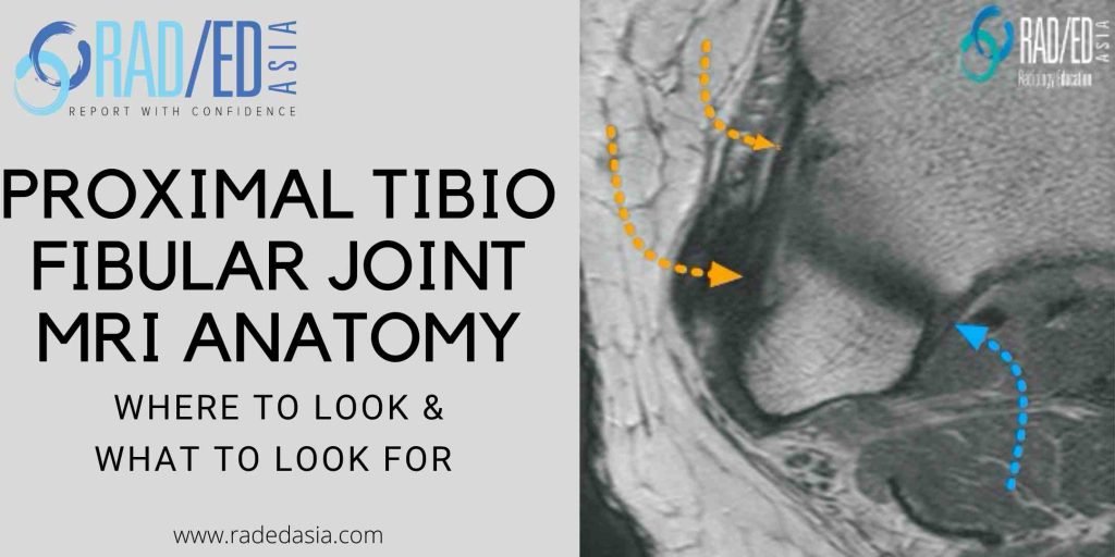
MRI KNEE ANATOMY: PROXIMAL TIBIOFIBULAR JOINT

PROXIMAL TIBIOFIBULAR JOINT MRI ANATOMY MRI PROXIMAL TIBIO FIBULAR JOINT ANATOMY The proximal tibiofibular joint is often overlooked when assessing a knee MRI. This is the first in a series on MRI of the proximal tibiofibular joint (PTFJ) beginning with the anatomy of the ligaments that stabilize the PTFJ. WHAT'S THE PROBLEM Often overlooked when …

MRI KNEE ANATOMY: PROXIMAL TIBIOFIBULAR JOINT Read More »

mri gleno humeral ligaments shoulder superior SGHL radiology education asia

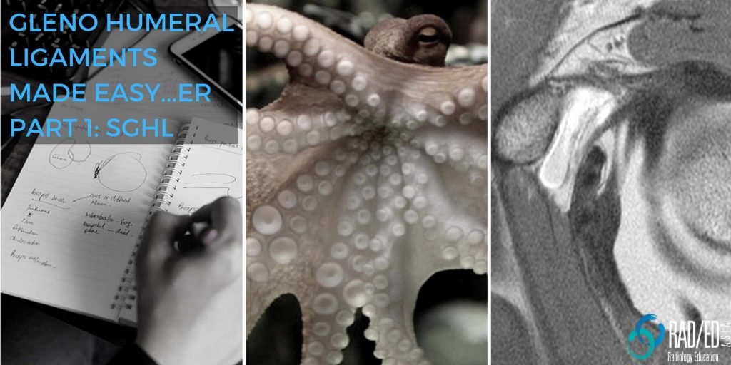
GLENOHUMERAL LIGAMENTS MRI ANATOMY: SUPERIOR GLENO HUMERAL LIGAMENT SGHL

SUPERIOR GLENOHUMERAL LIGAMENT SGHL SGHL: How to identify the Gleno Humeral Ligaments made Easy…er Identifying the gleno humeral ligaments on MRI can be challenging as they are small and on lower field strength scanners there is inadequate resolution to see them properly. However, if we understand the anatomy of the ligaments, it becomes easier to …

GLENOHUMERAL LIGAMENTS MRI ANATOMY: SUPERIOR GLENO HUMERAL LIGAMENT SGHL Read More »

proximal tibio fibular joint mri normal anatomy ligaments biceps femoris online radiology courses conference education asia

TUESDAY TIP: MRI OF THE PROXIMAL TIBIOFIBULAR JOINT (PART 2)

Today’s Tuesday Tip continues with the Proximal TibioFibular Joint. On MRI at the Proximal TibioFibular Joint ( PTFJ) one thing that can be confusing is the tibial attachment of the Biceps Femoris Tendon which can be mistaken for the AnteroSuperior TibioFibular Ligament. The Biceps Femoris tendon attaches predominantly to the fibula head but there is …

TUESDAY TIP: MRI OF THE PROXIMAL TIBIOFIBULAR JOINT (PART 2) Read More »

arcuate segond fracture fibular styloid radiology education asia tuesday tip

ARCUATE FRACTURE KNEE X-RAY, SEGOND FRACTURE, LATERAL KNEE FRACTURES

ARCUATE FRACTURE, SEGOND FRACTURE, LATERAL KNEE FRACTURES ARCUATE FRACTURE KNEE X-RAY , SEGOND FRACTURE, LATERAL FIBULAR FRACTURE This post looks at the 3 fractures (Segond Fracture, Arcuate Fracture and Lateral Fibular Fracture ) that can be seen on the lateral aspect of the knee x-ray which are indicative of underlying tendon or ligament involvement. The …

ARCUATE FRACTURE KNEE X-RAY, SEGOND FRACTURE, LATERAL KNEE FRACTURES Read More »

shoulder-cartilage-glenoid-dislocation

Rotator Cuff Tendon Tear on MRI : Dont miss the Leading Edge of Suprasinatus

Today’s Tuesday Tip is on How to find Supraspinatus Rotator Cuff tears on MRI. One of the easy to miss locations for a supraspinatus tendon tear is at the leading edge of the SST. In this video we look at what the leading edge is and how to find it.     Whats the Purpose …

Rotator Cuff Tendon Tear on MRI : Dont miss the Leading Edge of Suprasinatus Read More »

shoulder-cartilage-glenoid-dislocation

MRI GLENOID CARTILAGE MRI SHOULDER WHERE & WHAT TO LOOK FOR SHOULDER DISLOCATIONS

MRI GLENOID CARTILAGE GLENOID CARTILAGE MRI :What to look for and where to look for chondral damage in shoulder dislocations.   GLENOID CARTILAGE MRI:Glenoid cartilage damage can be seen with anterior and posterior shoulder dislocations on MRI and are part of the so called GLAD lesions.  Its important to first recognize the normal appearance of …

MRI GLENOID CARTILAGE MRI SHOULDER WHERE & WHAT TO LOOK FOR SHOULDER DISLOCATIONS Read More »

This site is intended for Medical Professionals only. Use of this site is governed by our Terms of Service and Privacy Statement which can be found by clicking on the links. Please accept before proceeding to the website.
error: Right Click not available.