proximal tibiofibular joint ligament anatomy mri

MRI KNEE ANATOMY: PROXIMAL TIBIOFIBULAR JOINT

MRI PROXIMAL TIBIO FIBULAR JOINT ANATOMY

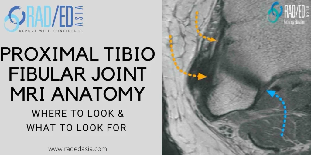
The proximal tibiofibular joint is often overlooked when assessing a knee MRI. This is the first in a series on MRI of the proximal tibiofibular joint (PTFJ) beginning with the anatomy of the ligaments that stabilize the PTFJ.

Often overlooked when assessing knee MRIs.

  • Joint stabilized by:
    1. Fibrous Capsule.
    2. Two ligaments reinforce capsule.


  • Also, fibula attachment of LCL, Biceps Femoris.
  • Runs obliquely from anterior fibula head to anterior lateral tibial condyle (Orange Arrow).


  • Normal MRI.

    1. Well defined, low signal, runs obliquely.
    2. Can be striated (2-3 bands).
    3. May be fused with tibial attachment of Biceps femoris.
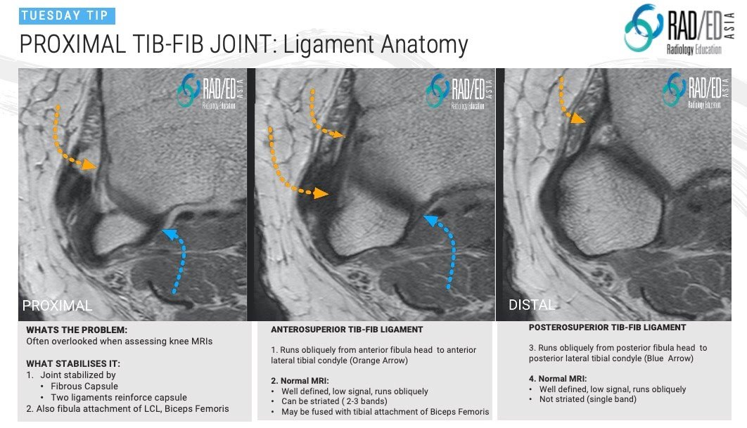
  • Runs obliquely from posterior fibula head to posterior lateral tibial condyle (Blue Arrow).


  • Normal MRI.

    1. Well defined, low signal, runs obliquely.
    2. Not striated (Single band).

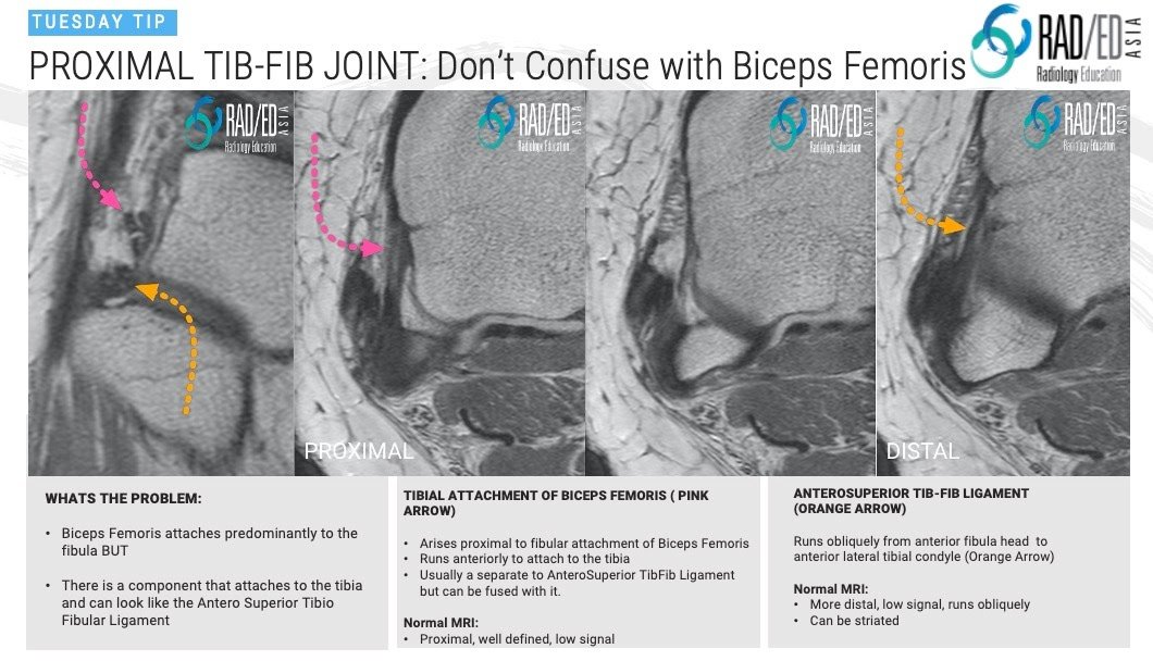
On MRI at the Proximal TibioFibular Joint ( PTFJ) one thing that can be confusing is the tibial attachment of the Biceps Femoris Tendon which can be mistaken for the AnteroSuperior TibioFibular Ligament.

The Biceps Femoris tendon attaches predominantly to the fibula head but there is a component that:

  1. Arises proximal to the fibula.
  2. Runs anteriorly and,
  3. Attaches to the lateral margin of the lateral tibial condyle and can look just like the antero-superior tibiofibular ligament.
  • Biceps femoris attaches predominantly to the fibula BUT,


  • There is a component that attaches to the tibia and can look like the Antero Superior Tibio Fibular Ligament.
  • Arises proximal to fibular attachment of Biceps femoris.
  • Runs anteriorly to attach to the tibia.
  • Usually a separate to Antero Superior Tib-Fib Ligament but can be fused with it.


Normal MRI.
Proximal, well defined, low signal.

Runs obliquely from anterior fibula head to anterior lateral tibial condyle (Orange Arrow).


Normal MRI.

  • More distal, low signal, runs obliquely.
  • Can be striated.

SO HOW DO YOU DIFFERENTIATE THE TWO?


  1. When you scroll on the axial scans from superior to inferior, the Biceps Femoris tendon is seen first (Pink arrows).
  2. There is usually a gap between the tendon and ligament (like in the image).
  3. The next structure you will see is the Ligament (Orange Arrows).
  4. So the first linear structure you see is the tendon (Pink arrows) and the more distal linear structure is the ligament (Orange arrow).
  5. Occasionally the two are fused and there will only be one linear structure.

Learn more about KNEE Imaging in our ONLINE or ONSITE
Guided MRI KNEE Mini-Fellowship.


More by clicking on the images below.

mri knee meniscus tears cysts pathologies images
KNEE
meniscal ossicles mri knee radiology education asia
KNEE

#kneemri #msk #mskmri #radedasia #sportsphysician #sportsmed #sportsmedicine #orthopaedic #chiropractic #orthopaedic #painphysician #physiotherapy #sportsinjury #rheumatology #arthritis #rheumatologist #physio #physiotherapist #radiology #mriknee #tibiofibular joint

#radedasia #mri #mskmri #radiology

This site is intended for Medical Professionals only. Use of this site is governed by our Terms of Service and Privacy Statement which can be found by clicking on the links. Please accept before proceeding to the website.
error: Right Click not available.