Identifying the gleno humeral ligaments on MRI can be challenging as they are small and on lower field strength scanners there is inadequate resolution to see them properly.
However, if we understand the anatomy of the ligaments, it becomes easier to know where to look even if we can't directly identify the ligament.
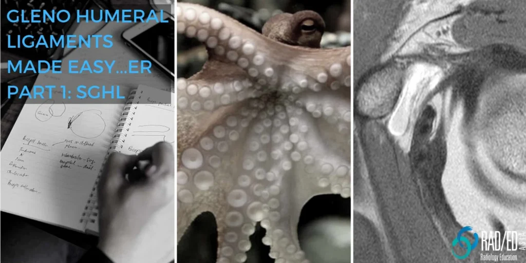
This post is an adaptation of a talk given at Radiology Asia in Singapore titled Gleno Humeral Ligaments made Easy…er.![]()
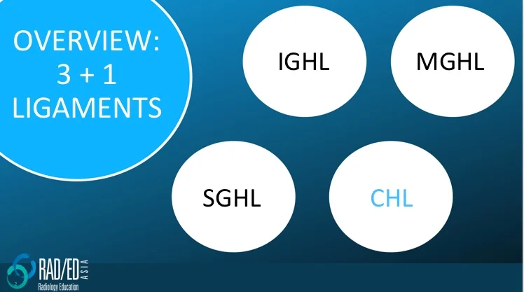
1. SGHL: Superior GlenoHumeral Ligament
2. MGHL: Middle GlenoHumeral Ligament
3. IGHL: Inferior GlenoHumeral Ligament
4. The Coraco Humeral Ligament (CHL) is extra capsular and is not part of the Glenohumeral ligaments, but we will also look at as it helps to identify the SGHL and it also fuses with the joint capsule at the rotator interval. (Click here for more on the CHL)
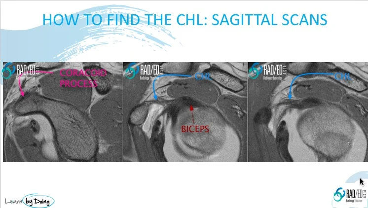
- The CHL is best seen on sagittal scans and the Coracoid Process is the anatomical marker we use to identify it.
- Start medially find the coracoid process (Pink arrow).
- Scroll laterally and the first horizontal structure you see going from coracoid over to the biceps insertion is the coraco humeral ligament.
![]()
The ligaments arise in an orderly fashion and using analogies often helps to understand anatomy. On sagittal scans the ligaments have the appearance of tentacles of an octopus radiating from the biceps insertion on the supra glenoid tubercle.
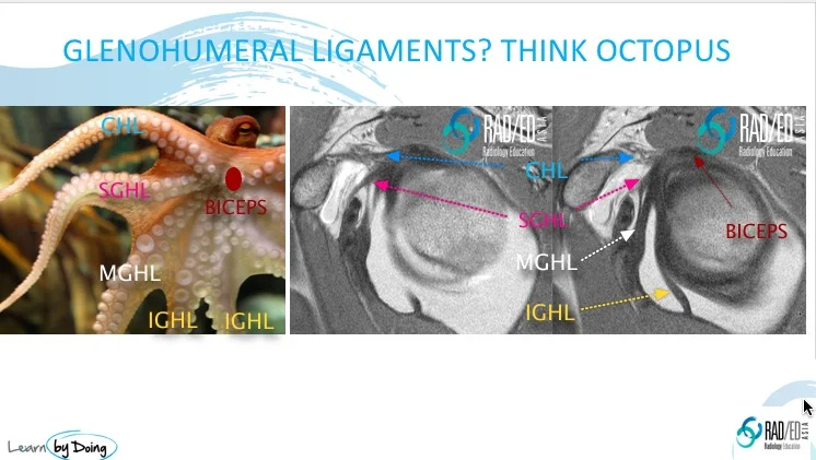
Image Above:
- After having identified the CHL, the next structure that you see immediately below it on sagittal scans is the SGHL (Pink arrow).
- The Biceps insertion (Crimson arrow) is our anatomical marker for the SGHL.
- SGHL (Pink arrow) arises at or just below the biceps insertion.

Image Above:
Continuing to scroll laterally, the CHL (Blue arrow) and the SGHL (Pink arrow) start to approximate each other and fuse with the capsule (Yellow arrow) to form one structure, the Rotator Interval Capsule.![]()
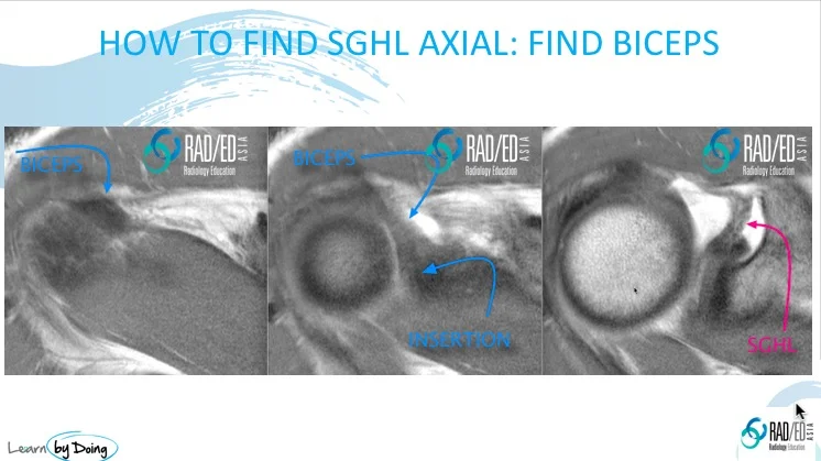
- On axial scans our anatomical marker is the biceps tendon.
- Start on the highest axial scan and scroll inferiorly.
- Find biceps tendon (Blue arrow) arching over the anterior superior humeral head and identify its insertion.
- At the level of the insertion or on 1 slice below, a linear structure passing anteriorly to the capsule is the SGHL (Pink arrow).
![]()
The three main Glenohumeral ligaments are the Superior GlenoHumeral Ligament (SGHL), Middle GlenoHumeral Ligament (MGHL), and Inferior GlenoHumeral Ligament (IGHL).
![]()
The extra capsular ligament that is not part of the Glenohumeral ligaments is the Coraco Humeral Ligament (CHL).
![]()
The CHL is best seen on sagittal scans and the Coracoid Process is the anatomical marker used to identify it.
![]()
The ligaments on sagittal scans have the appearance of tentacles of an octopus radiating from the biceps insertion on the supra glenoid tubercle.
![]()
The Biceps insertion is the anatomical marker used to identify the SGHL on sagittal scans.
![]()
The structure that is formed when the CHL and SGHL fuse with the capsule on lateral scrolling in sagittal scans is the Rotator Interval Capsule.
![]()