mri knee meniscus root tear anatomy radedasia

KNEE MRI MENISCUS ROOT TEAR: WHERE TO LOOK

Each meniscus has an anterior and posterior root and there are four meniscal roots but we will focus on the Posterior root of the medial meniscus as this is the root most commonly torn. In this post look at the imaging anatomy of the medial meniscus root on MRI.

  • The meniscal root is a small portion of the meniscus that attaches and anchors the anterior and posterior horns of the meniscus to the intercondylar fossa or eminence of the tibia.


  • Its usually the posterior root of the meniscus that is torn and medial more than lateral meniscus.

It is the component of the meniscus between its tibial attachment and the point on the tibial margin where cartilage ends.


mri knee meniscus root post horn medial anatomy radedasia


Image Above: Red arrows demonstrate the medial meniscus root. It commences just lateral to the apex of the medial tibial plateau (Yellow arrow). Green arrow and dashed green line demonstrate where cartilage of medial tibial plateau ceases which is another marker for the beginning of the meniscal root.

The posterior horn root of the medial meniscus attaches immediately anterior to the PCL.


mri knee post horn medial meniscus root radedasia


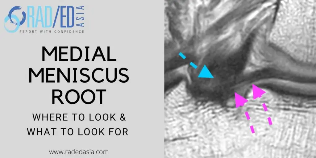
Image above: Coronal scans Pink arrows demonstrates attachment of medial meniscus root to tibia anterior to PCL (Blue arrows).

On the sagittal scans the root commences as a triangular structure  at the posterior horn/root junction medially (Image A) and progressively becomes more ovoid as it inserts into the tibia laterally (Image D). It inserts anterior to the PCL.


knee mri meniscus root tear anatomy


Sagittal Images above: Yellow arrow demonstrates medial meniscus posterior horn and meniscal root (Yellow arrow). Note how meniscal root extends to the level of the PCL (Green arrow).

A tear of the meniscal root results in significant dysfunction of the meniscus because:

  • The meniscal root anchors the meniscus and prevents its being extruded.


  • This enables the meniscus to generate hoop stress which transfers load from the femur to the tibia without damage to underlying cartilage.


  • The effect of a meniscal root tear is equivalent to having a total meniscectomy in terms of function and increases risk of damage to cartilage.
  • Tears are best seen on the coronal images and then confirmed on the sagittal scans.


  • The root of the posterior horn medial meniscus attached immediately anterior to the PCL. On the sagittal scan you should see the posterior horn on every sagittal image until the PCL.


  • There should be no fluid between the PCL and the meniscal root on the coronal scans.


  • We will look at the MRI appearance of tears in a subsequent post.

We look at all of these topics in more detail in our KNEE MRI
Guided Mini Fellowships.

Click on the image below for more information.

  • Join our WhatsApp Group for regular educational posts. Message “JOIN GROUP” to +6594882623 (your name and number remain private and cannot be seen by others).

  • Get our weekly email with all our educational posts: https://bit.ly/whathappendthisweek
spine-imaging-course-pain-management-sports-medicine-orthopaedic-specialists-ct-mri
ONLINE MINI-FELLOWSHIPS
radiology knee segond arcuate lateral fibular fracture xray mri knee radedasia
KNEE
iliotibial band mri anatomy syndrome trauma mri knee radedasia
KNEE

#radiology #radiologist #radiologia #mri #kneemri #mskmri #roottear #mskrad #mskradiology #imaging #frcr #sportsmed #radiologyresident #foamrad #ortho #ct #radiologystudent #trauma #radedasia #radiologycme #radiologyeducation #radiologycases #rheumatology #arthritis #painphysician #physiotherapy

#radedasia #mri #mskmri #radiology

Leave a Comment

This site is intended for Medical Professionals only. Use of this site is governed by our Terms of Service and Privacy Statement which can be found by clicking on the links. Please accept before proceeding to the website.
error: Right Click not available.