acromial spur mri shoulder radiology education asia

ACROMION / ACROMIAL SPURS ON MRI: WHERE TO LOOK AND WHAT TO LOOK FOR

ACROMION SPURS ON MRI: 3 APPEARANCES ON MRI


Acromial / Acromion spurs can be quite small and difficult to visualize on MRI.


They are enthesophytes that arise from traction at the attachment sites of the Coraco Acromial Ligament and Deltoid.


This post looks at the three locations and most common appearances of spurs arising from the lateral aspect of the acromion.

WHAT’S THE NORMAL APPEARANCE OF THE LATERAL ACROMION

 
  • The normal lateral tip of the acromion has a smooth, ovoid contour (blue arrow).
  • The under-surface of the acromion is also smooth and continuous (pink arrow) with no step or ledge.
  • subacromial spur mri shoulder normal acromion radiology education asia 1

    Beak like spurs form on the lateral tip of the acromion. They can project either:

    • Laterally and parallel to the acromion
    • Or project inferiorly from the tip of the acromion.


    subacromial spur mri shoulder radiology education asia 4


    Image Above: Beak like spur (blue arrows). 

    • The rounded appearance of the lateral margin of the acromion is lost.
    • The cortex and marrow of the acromion are continuous around and into the spur.

    These are seen on sagittal scans as they project anteriorly from the acromion and occur at the attachment site of the coraco acromial ligament.

    acromial spur anterior beak like mri shoulder anterior radiology education asia


    Image Above: 

    • Normal rounded appearance of anterior acromion margin (Pink arrow) at the attachment of the coraco acromial ligament (Blue arrow).

    • Anterior beak like spur (yellow arrow) with loss of round anterior acromial margin.

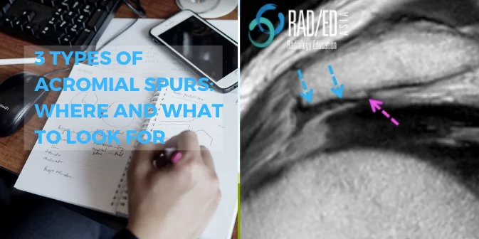
    • These spurs are more broad based and arise from the under-surface of the lateral acromion (blue arrows below).


    • The Coraco Acromial ligament attaches to the under-surface and a broad traction enthesophyte arises at the insertion site.


    • The normal under surface is smooth and continuous with the tip without a step in the cortex.


    • When a spur forms there is a step in the cortex (Pink arrows below) giving it a broad shelf like appearance.


    • The cortex is continuous around and the spur and marrow extends into the spur.


    sub acromial spur mri heel shoulder radiology education asia

    • Join our WhatsApp Group for regular educational posts. Message “JOIN GROUP” to +6594882623 (your name and number remain private and cannot be seen by others).

    • Get our weekly email with all our educational posts: https://bit.ly/whathappendthisweek

    #radedasia #mri #mskmri #radiology

    This site is intended for Medical Professionals only. Use of this site is governed by our Terms of Service and Privacy Statement which can be found by clicking on the links. Please accept before proceeding to the website.
    error: Right Click not available.