hip labrum ossification mri

HIP LABRUM OSSIFICATION MRI: LABRAL ABNORMALITIES MORE THAN JUST TEARS PART 2

Acetabular labrum ossification is not common and I have been able to find only a few reports on it. The cases we have seen mostly have an underlying dysplastic, elongated labrum however there are reports of it occurring in a normal labrum. So what does it look like? Having learnt the hard way by missing them by looking only at the MRI, I find the best place to start is with the plain X-ray as it can be overlooked if assessing just the MRI.

hip-labrum-ossification-radedasia
 
Hip-labrum-ossifiction-xray-radedasia

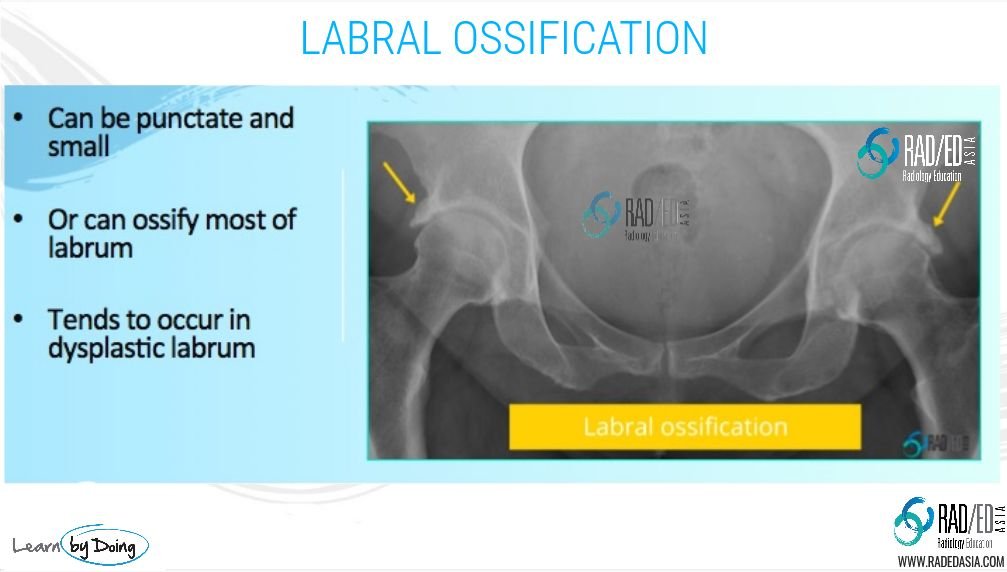
Image 2 Above: Bilateral labral ossification in the same patient. Labrum is diffusely ossified. Ossification follows the shape of the labrum. MRI below.

hip-labrum-ossification-mri-radedasia

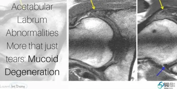
Image 3 Above: MRI for patient in Image 2. On the PD and PDFS scans ossification follows same signal as adjacent bone. Labrum is elongated and dysplastic.

hip-labrum-ossification-mri-rea-radedasia

Image 4 above: PD and PDFS ossification of the labrum centered on the labrum and follows labrum shape. Different patient to Image 2 and 3.

Learn more about HIP Imaging in our ONLINE
Guided MRI HIP Mini-Fellowship.


More by clicking on the images below.

#mrilabrum #labrumossification #labrummucoiddegeneration #radiology #radedasia #mri #hipmri #radiologyeducation #radiologycases #radiologist #radiologycme #radiologycpd #medicalimaging #imaging #radcme #rheumatology #arthritis #rheumatologist #orthopaedic #painphysician #podiatry #podiatrist #physiotherapy #sportsmed #orthopaedic #mskmri

#radedasia #mri #mskmri #radiology

This site is intended for Medical Professionals only. Use of this site is governed by our Terms of Service and Privacy Statement which can be found by clicking on the links. Please accept before proceeding to the website.
error: Right Click not available.