
Extensive synovitis and ACJ erosions from Rheumatoid arthritis. Having synovitis in multiple locations (even if there are no erosions) should raise the possibility of an inflammatory abnormality.
Thank you to Dr Joe Thomas (Rheumatologist) for the case.
![]()
Image 1: The first image demonstrates marked synovitis in the joint and the biceps tendon sheath. Notice the different types of appearance of synovitis in the posterior joint (Pink arrow), Anterior joint (Yellow arrow) and the Biceps tendon sheath (Yellow arrow). All are intermediate signal on T2FS.
Image 2: Synovitis also present in the subacromial subdeltoid bursa (Blue arrows).
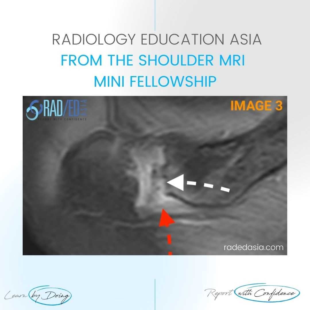
Image 3: AC Joint Synovitis (Red arrow) and Erosions (White arrow).
![]()
 This post has been made in conjunction with Dr Joe Thomas, a Senior Consultant Rheumatologist at Aster Hospital in Kochi India, who has a vast amount of clinical experience. He also has a very strong interest in Imaging of Arthropathies and has joined us to bring a clinical perspective to the imaging and to advise on what rheumatologists want when we report their referrals.


