Distal biceps tendon anatomy with Long and Short Heads, tendonosis, tear, and rupture that can affect one head only.

Stay tuned on new Fellowships and learnings

MRI DISTAL BICEPS TENDON: LONG vs SHORT HEAD PATHOLOGY — WHAT TO LOOK FOR

MRI DISTAL BICEPS TENDON: LONG VS SHORT HEAD PATHOLOGY — WHAT TO LOOK FOR

The distal biceps tendon has two heads — and one can tear while the other stays normal

  • The distal biceps tendon is made up of two separate tendons — the Long Head and the Short Head of Biceps — that fuse together before inserting onto the radial (bicipital) tuberosity.
  • Tendonosis, tears, or rupture can affect the tendon of one head only, with the other head spared.
  • Diagnosing these abnormalities starts with knowing the normal anatomy of the distal biceps tendon.
  • The MRI appearance of tendonosis, tears, and rupture in this tendon follows the same general appearance as tendon pathology elsewhere in the body.
DISTAL BICEPS TENDON ANATOMY
biceps tendon anatomy mri radiology education asia radedasia biceps tendon distal normal elbow mri radiology education asia radedasia
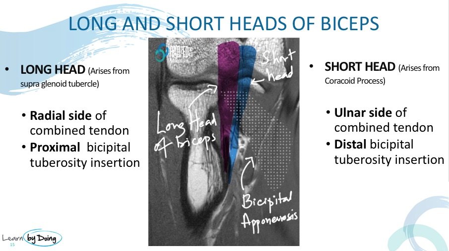
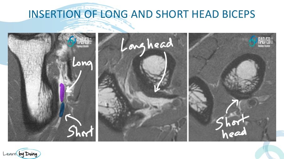
  • The tendon of the Long Head of Biceps lies on the radial side of the combined tendon, with a proximal insertion on the radial (bicipital) tuberosity.
  • The tendon of the Short Head of Biceps lies on the ulnar side of the combined tendon, with a distal insertion on the radial (bicipital) tuberosity.
  • On axial scans, the Long Head (purple arrows), Short Head (blue arrows), and combined tendon (yellow arrows) can be identified as separate structures.
biceps tendon normal anatomy mri elbow radiology education asia radedasia

On the coronal image, the Long Head inserts more superiorly (purple region) than the Short Head (blue region).

MRI APPEARANCE OF DISTAL BICEPS TENDON ABNORMALITIES
biceps tendonosis mri long head radiology education asia radedasia
  • Tendonosis predominantly involves the Long Head, with the Short Head less involved.
  • On axial imaging, a difference in signal is seen between the Long and Short Heads at their respective levels.
biceps tendon mri tear short head radiology education asia radedasia

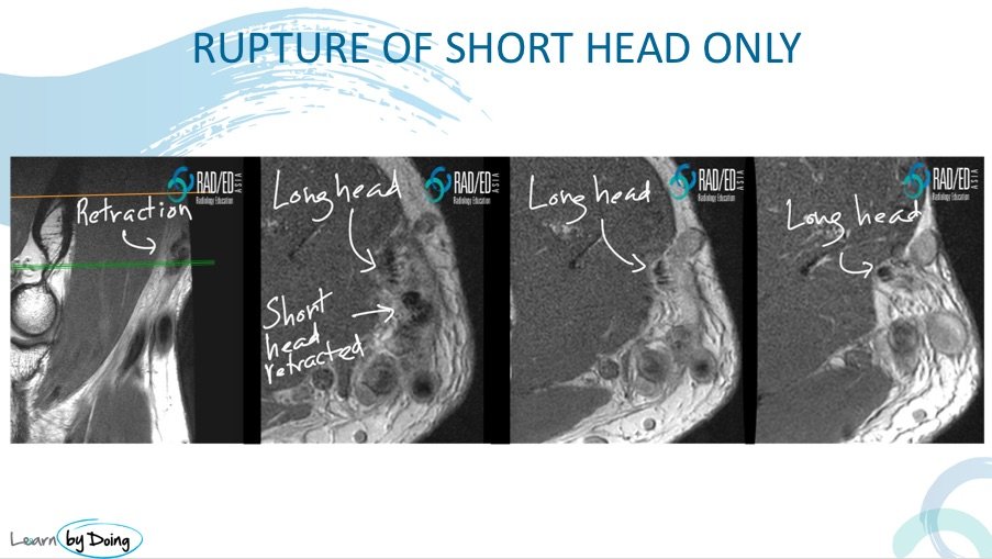
Severe tendonosis and tear of the Short Head of Biceps can occur with sparing of the Long Head.

biceps tendon rupture tear mri radiology education asia radedasia
  • When the Short Head ruptures and retracts, only a single tendon is seen on axial images where two tendons should normally be present.
  • The Long Head can remain attached to the radial tuberosity even when the Short Head has ruptured.
KEY POINT
  • The distal biceps tendon is formed by two separate tendons — the Long Head (radial side, proximal insertion) and the Short Head (ulnar side, distal insertion) — that fuse before inserting on the radial tuberosity.
  • Tendonosis, tears, and rupture can selectively affect one head while sparing the other, so assess the Long and Short Heads independently.
  • On axial images, look for asymmetric signal between the two heads, or a missing tendon, to identify one-head-only pathology.
  • A ruptured and retracted head may leave only a single tendon visible on axial images, while the unaffected head remains normally attached.
TEST YOURSELF ON SOME COMMON & FREQUENTLY ASKED QUESTIONS

Yes. Tendonosis, tears, or rupture can affect the tendon of one head only, with the other head spared.

The Long Head lies on the radial side of the combined tendon and has a proximal insertion on the radial (bicipital) tuberosity.

The Short Head lies on the ulnar side of the combined tendon and has a distal insertion on the radial (bicipital) tuberosity.

The Long Head inserts more superiorly than the Short Head.

Only a single tendon is seen on axial images where two tendons should normally be present, while the Long Head may remain attached to the radial tuberosity.

Our CPD & Learning Partners

OUR OTHER POPULAR POSTS ON GLENOHUMERAL LIGAMENTS: CLICK ON THE IMAGE BELOW

Stay tuned on new Fellowships and learnings

error: Content is protected !!

Suscríbase a nuestro boletín

Esté atento a los nuevos lanzamientos y aprendizajes de las Fellowships

Fellowships

Mejor que las conferencias

Únase a nuestra comunidad de WhatsApp

Ayudamos a los radiólogos a aprender
haciendo e informa con confianza

Suscríbase a nuestro boletín

Esté atento a los nuevos lanzamientos y aprendizajes de las Fellowships

Fellowships

Mejor que las conferencias

Únase a nuestra comunidad de WhatsApp

Ayudamos a los radiólogos a aprender
haciendo e informa con confianza

Este sitio está destinado exclusivamente a las profesiones médicas. El uso de este sitio se rige por nuestras Condiciones de servicio y Declaración de privacidad, que puede consultar haciendo clic en los enlaces. Por favor, acéptelas antes de continuar en el sitio web.