
Este sitio está destinado exclusivamente a las profesiones médicas. El uso de este sitio se rige por nuestras Condiciones de servicio y Declaración de privacidad, que puede consultar haciendo clic en los enlaces. Por favor, acéptelas antes de continuar en el sitio web.
10
10 years of RAD/ED — from one room in KL to thousands worldwide. Thank you.
MRI SYNOVITIS KNEE: THE MANY APPEARANCES — WHAT TO LOOK FOR
Synovitis can take many forms on MRI — recognise the pattern once and you will spot it in any joint
- Synovitis is usually generalised and associated with an effusion, but can also be localised to a portion of the joint without an effusion.
- Inflammatory and infective synovitis cannot be differentiated on MRI — contrast will not help, as all forms of synovitis will enhance.
- The morphological appearances below are consistent across joints. Learn to recognise them in the knee and you will apply the same pattern elsewhere.
MRI APPEARANCES OF SYNOVITIS
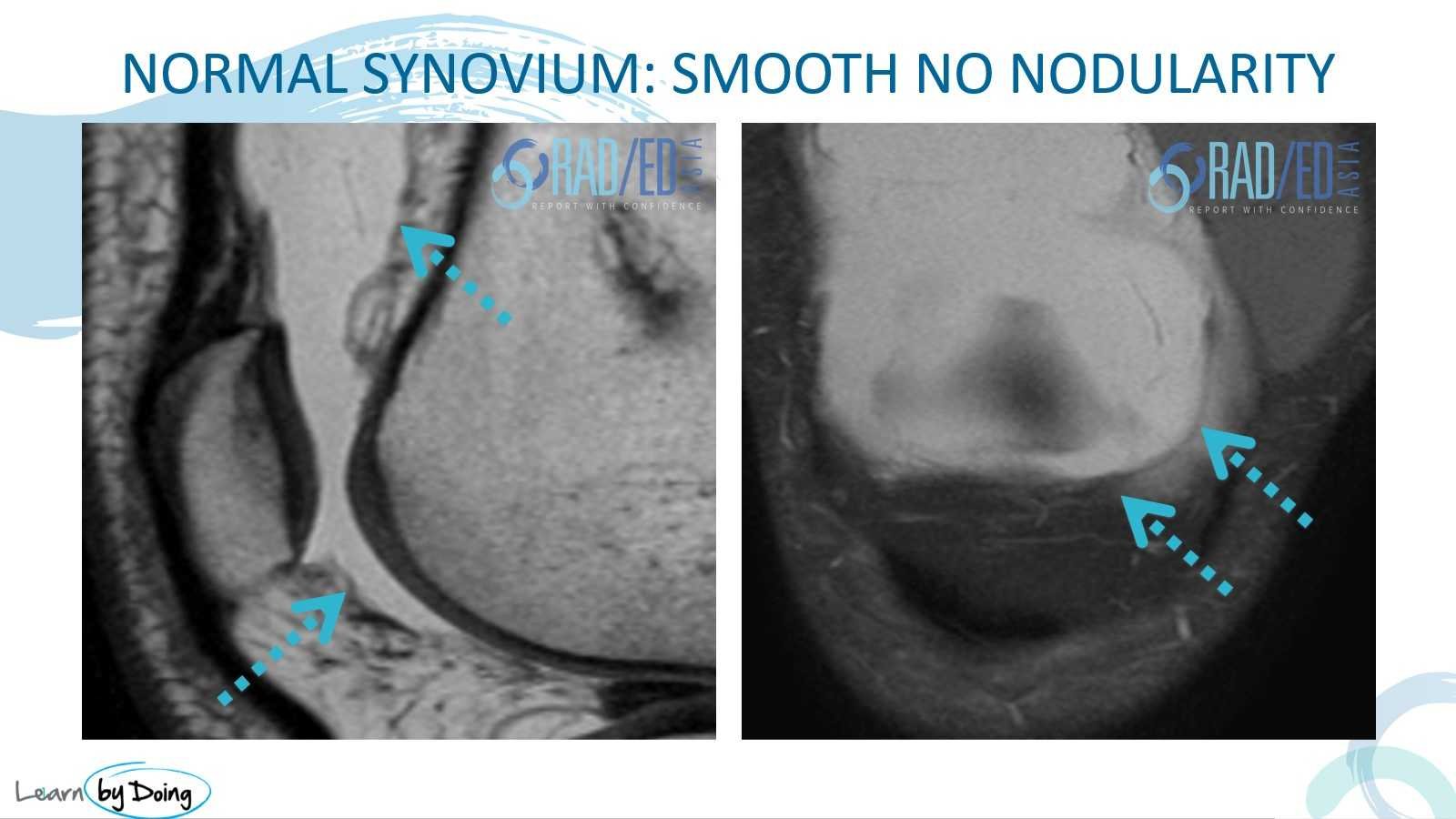
- Normally you cannot differentiate synovium from capsule — what we see is a thin, low-signal line.
- Best assessed on T2/PD sequences.
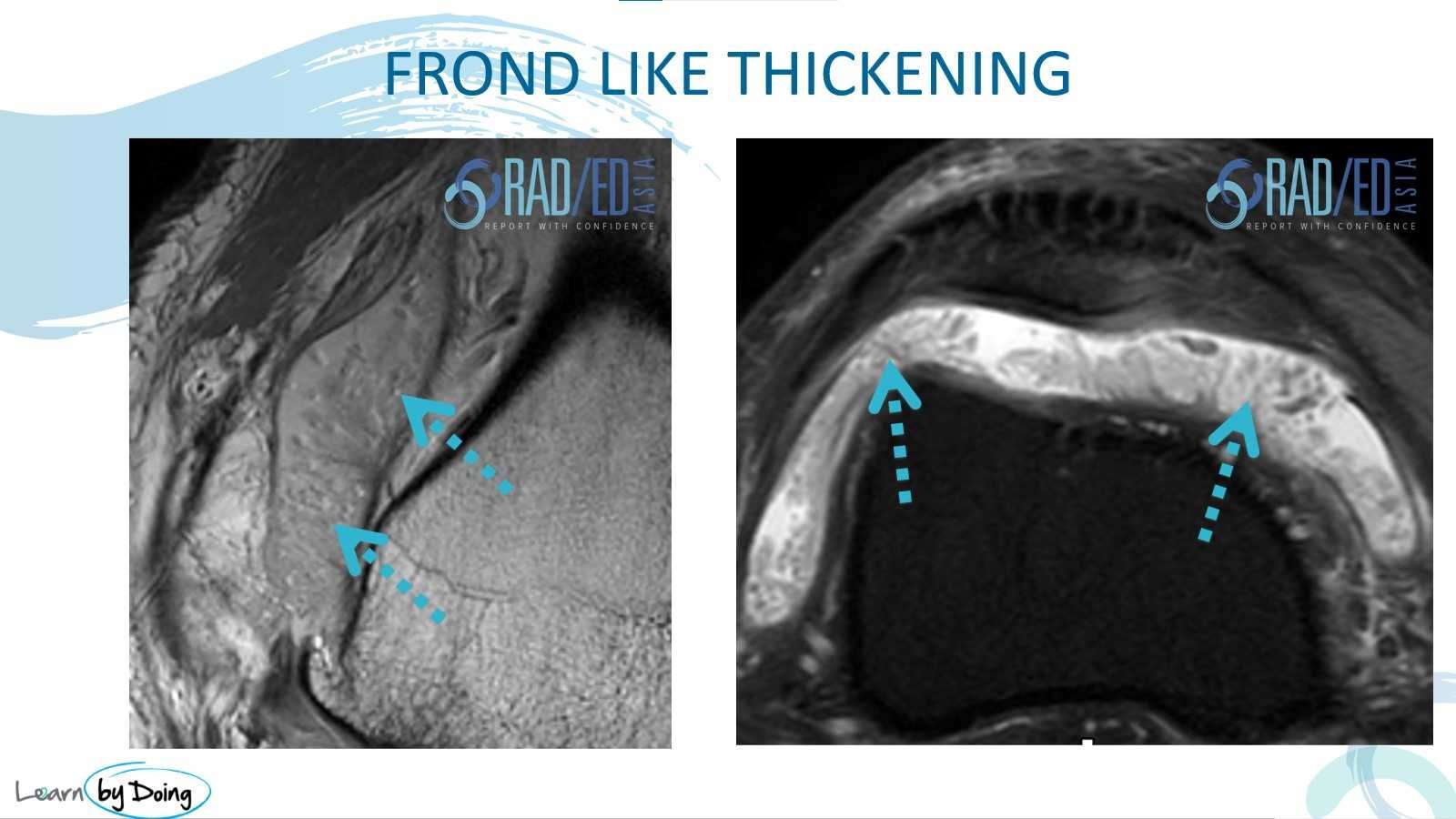
- The most common appearance — thin, fibrillary morphology, intermediate signal on T2 and PD.
- Much easier to detect on T2/PD than on T1.
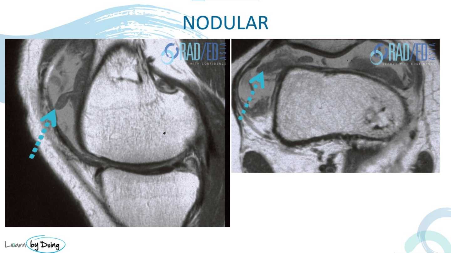
- Thicker appearance than frond-like synovitis — can be elongated or rounded and nodular.
- Nodular synovitis should raise the possibility of PVNS (pigmented villonodular synovitis).
More extensive synovitis with a solid, sheet-like morphology rather than discrete fronds or nodules.
- Similar to the sheet-like appearance but lines the joint capsule.
- Most commonly seen post ACL repair — represents a form of arthrofibrosis.
- Usually low signal on PD/T2.
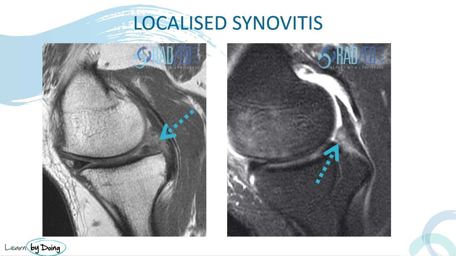
- Intermediate-signal synovitis confined to a specific area — commonly posterior to the meniscus.
- Often associated with a meniscal tear, which is the likely cause of the localised synovitis in that region.
KEY POINT
- Synovitis presents as frond-like (most common), nodular, sheet-like, confluent capsular thickening, or localised — each with a distinct MRI morphology.
- All types are best assessed on T2/PD sequences; T1 is much less sensitive.
- Nodular synovitis should raise the possibility of PVNS.
- Confluent capsular thickening is a form of arthrofibrosis, most often seen post ACL repair, and is typically low signal on PD/T2.
- Inflammatory and infective synovitis cannot be distinguished on MRI — contrast enhances all forms equally and does not assist differentiation.
TEST YOURSELF ON SOME COMMON & FREQUENTLY ASKED QUESTIONS
T2 and PD sequences. Synovitis is intermediate in signal and much easier to detect on T2/PD than on T1.
No. Inflammatory and infective synovitis cannot be differentiated on MRI. Contrast does not help — all forms of synovitis enhance.
PVNS (pigmented villonodular synovitis). Nodular synovitis — particularly if elongated or rounded — should raise this possibility.
A form of arthrofibrosis in which thickened, low-signal tissue lines the joint capsule. Most commonly seen post ACL repair.

- Join our WhatsApp RadEdAsia community for regular educational posts at this link: https://bit.ly/radedasiacommunity
- Get our weekly email with all our educational posts: https://bit.ly/whathappendthisweek