
MRI synovitis of the knee showing varied appearances and key imaging findings for radiologists.

Stay tuned on new Fellowships and learnings

SYNOVITIS MRI FINDINGS KNEE WHAT DOES IT LOOK LIKE

10

10 years of RAD/ED — from one room in KL to thousands worldwide. Thank you.

MRI SYNOVITIS KNEE: THE MANY APPEARANCES — WHAT TO LOOK FOR

Synovitis can take many forms on MRI — recognise the pattern once and you will spot it in any joint
  • Synovitis is usually generalised and associated with an effusion, but can also be localised to a portion of the joint without an effusion.
  • Inflammatory and infective synovitis cannot be differentiated on MRI — contrast will not help, as all forms of synovitis will enhance.
  • The morphological appearances below are consistent across joints. Learn to recognise them in the knee and you will apply the same pattern elsewhere.
MRI APPEARANCES OF SYNOVITIS
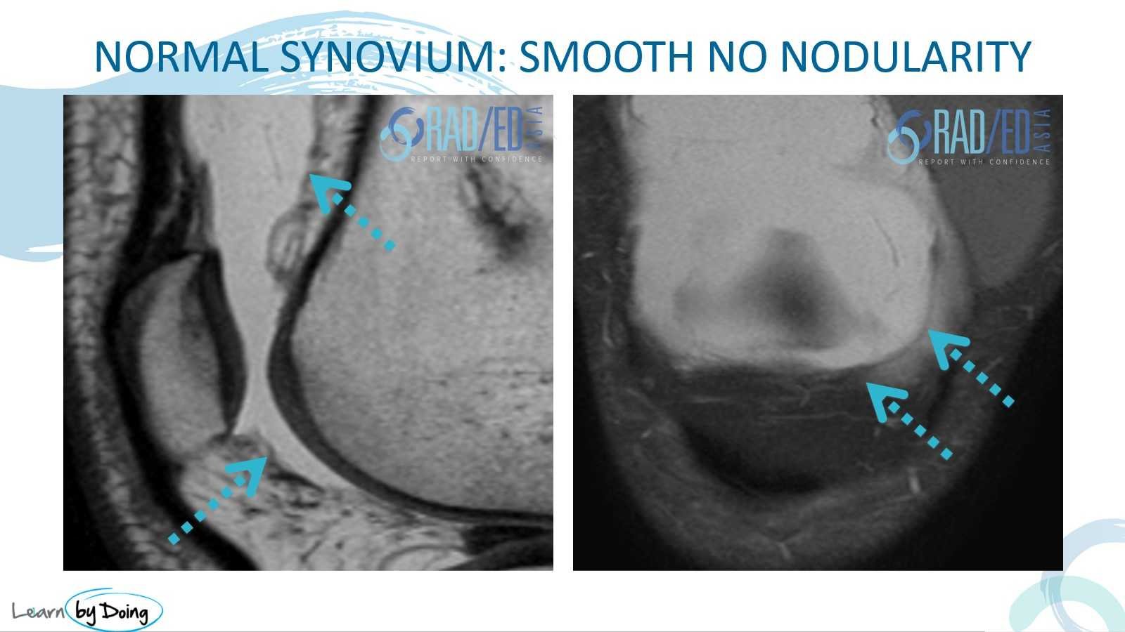
mri synovitis knee normal synovium radedasia
  • Normally you cannot differentiate synovium from capsule — what we see is a thin, low-signal line.
  • Best assessed on T2/PD sequences.
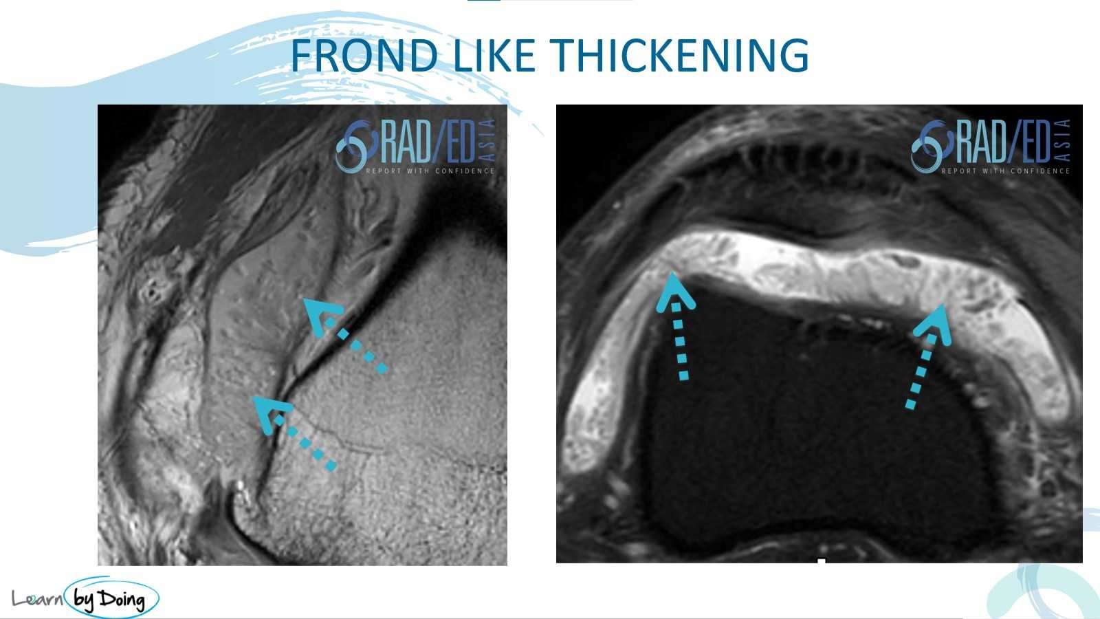
mri synovitis knee frond like radedasia
  • The most common appearance — thin, fibrillary morphology, intermediate signal on T2 and PD.
  • Much easier to detect on T2/PD than on T1.
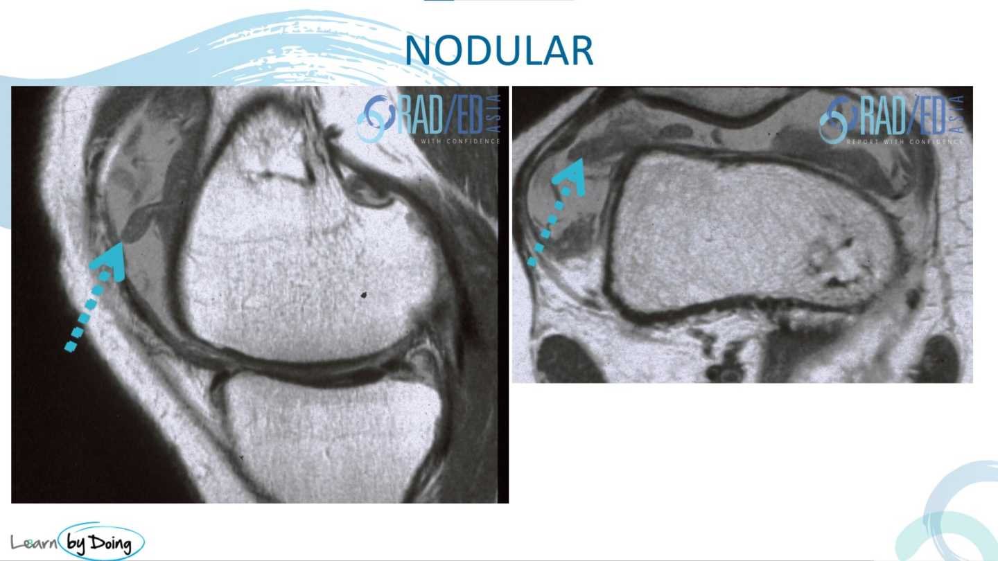
mri synovitis knee nodular radedasia
  • Thicker appearance than frond-like synovitis — can be elongated or rounded and nodular.
  • Nodular synovitis should raise the possibility of PVNS (pigmented villonodular synovitis).
mri synovitis knee sheet like thickening radedasia

More extensive synovitis with a solid, sheet-like morphology rather than discrete fronds or nodules.

mri synovitis knee confluent capsular thickening radedasia
  • Similar to the sheet-like appearance but lines the joint capsule.
  • Most commonly seen post ACL repair — represents a form of arthrofibrosis.
  • Usually low signal on PD/T2.
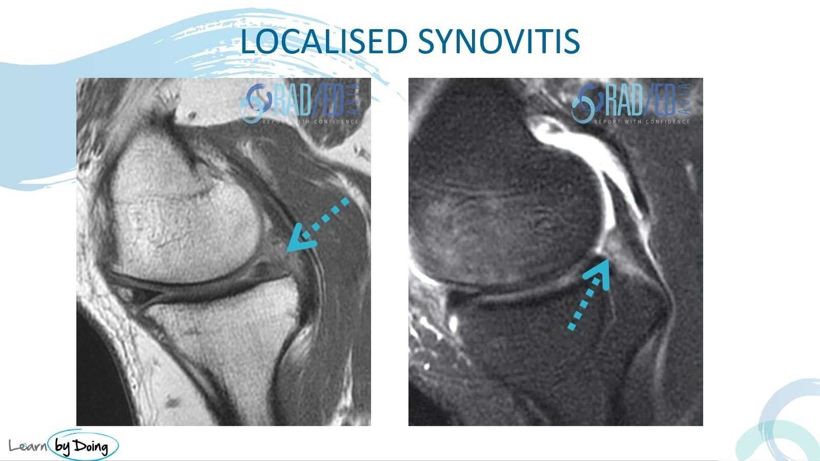
mri synovitis knee localised radedasia
  • Intermediate-signal synovitis confined to a specific area — commonly posterior to the meniscus.
  • Often associated with a meniscal tear, which is the likely cause of the localised synovitis in that region.
KEY POINT
  • Synovitis presents as frond-like (most common), nodular, sheet-like, confluent capsular thickening, or localised — each with a distinct MRI morphology.
  • All types are best assessed on T2/PD sequences; T1 is much less sensitive.
  • Nodular synovitis should raise the possibility of PVNS.
  • Confluent capsular thickening is a form of arthrofibrosis, most often seen post ACL repair, and is typically low signal on PD/T2.
  • Inflammatory and infective synovitis cannot be distinguished on MRI — contrast enhances all forms equally and does not assist differentiation.
TEST YOURSELF ON SOME COMMON & FREQUENTLY ASKED QUESTIONS

T2 and PD sequences. Synovitis is intermediate in signal and much easier to detect on T2/PD than on T1.

No. Inflammatory and infective synovitis cannot be differentiated on MRI. Contrast does not help — all forms of synovitis enhance.

PVNS (pigmented villonodular synovitis). Nodular synovitis — particularly if elongated or rounded — should raise this possibility.

A form of arthrofibrosis in which thickened, low-signal tissue lines the joint capsule. Most commonly seen post ACL repair.


Our CPD & Learning Partners

OUR POPULAR POSTS ON MENISCUS: CLICK ON THE IMAGES BELOW

meniscal tear radiology mri knee radedasia
KNEE
mri knee meniscus extrusion tear root
KNEE

Stay tuned on new Fellowships and learnings

error: Content is protected !!

Join our Newsletter

Stay tuned on new
Mini-Fellowships launches and learnings

Mini-Fellowships

Better Than Conferences

Join our WhatsApp Community

We help radiologists learn
by doing and report with confidence

This site is intended for Medical Professions only. Use of this site is governed by our Terms of Service and Privacy Statement which can be found by clicking on the links. Please accept before proceeding to the website.