Este sitio está destinado exclusivamente a las profesiones médicas. El uso de este sitio se rige por nuestras Condiciones de servicio y Declaración de privacidad, que puede consultar haciendo clic en los enlaces. Por favor, acéptelas antes de continuar en el sitio web.

CALCANEOFIBULAR LIGAMENT (CFL) TEAR MRI
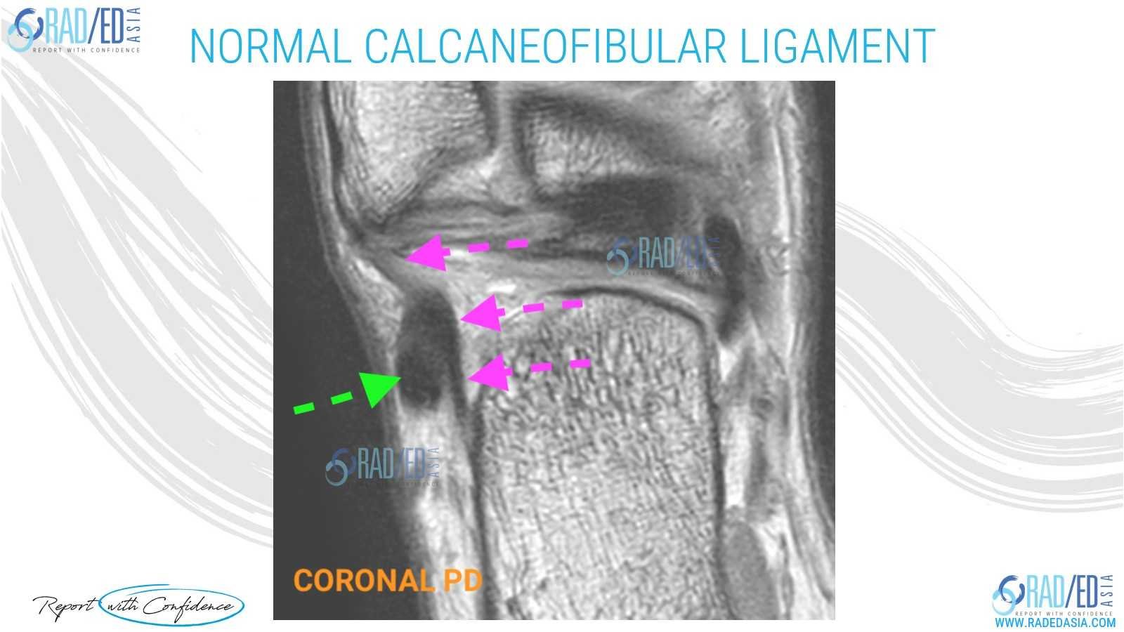
CALCANEOFIBULAR LIGAMENT (CFL) TEAR NORMAL MRI
- The Calcaneofibular ligament is a small lateral ankle ligament that is often torn.
- Its normal MRI appearance is a thin low signal structure (Pink arrows) that extends from the calcaneum to the fibula and passes deep to the peroneal tendons (Green arrows).

CALCANEOFIBULAR LIGAMENT (CFL) TEAR MRI ANKLE: WHAT TO LOOK FOR
The Calcaneofibular ligament is a small lateral ankle ligament that is often torn.
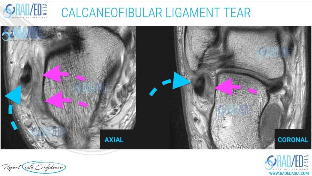
Here is what to look for when assessing for an acute tear of the Calcaneofibular ligament on MRI.
- Thickened, hyper-intense and ill defined calcaneofibular ligament (Pink arrows).
- This can involve the entire ligament or a portion of it.
- The anatomical landmark to find the CFL are the Peroneal tendons (Blue arrows) with the CFL lying deep to it between the peroneal tendons and calcaneum.
CALCANEOFIBULAR LIGAMENT (CFL) TEAR MRI ANKLE: HOW TO REPORT IT

TEST YOURSELF ON SOME COMMON & FREQUENTLY ASKED QUESTIONS
WHAT IS THE NORMAL MRI APPEARANCE OF THE CALCANEOFIBULAR LIGAMENT:
HOW DOES AN ACUTE HIGH-GRADE TEAR OF THE CALCANEOFIBULAR LIGAMENT APPEAR ON AN MRI?
WHAT ANATOMICAL LANDMARK CAN HELP LOCATE THE CALCANEOFIBULAR LIGAMENT ON AN MRI?
Our CPD & Learning Partners
Learn more about this condition & how best to report it in more detail in our Online Guided ANKLE or FOOT & TOE MSK MRI Mini Fellowship.
More by clicking on the images below
- Join our WhatsApp RadEdAsia community for regular educational posts at this link: https://bit.ly/radedasiacommunity
- Get our weekly email with all our educational posts: https://bit.ly/whathappendthisweek


