
ANKLE
The Calcaneofibular ligament is a small lateral ankle ligament that is often torn.
Here is what to look for when assessing for an acute tear of the Calcaneofibular ligament on MRI.
![]()

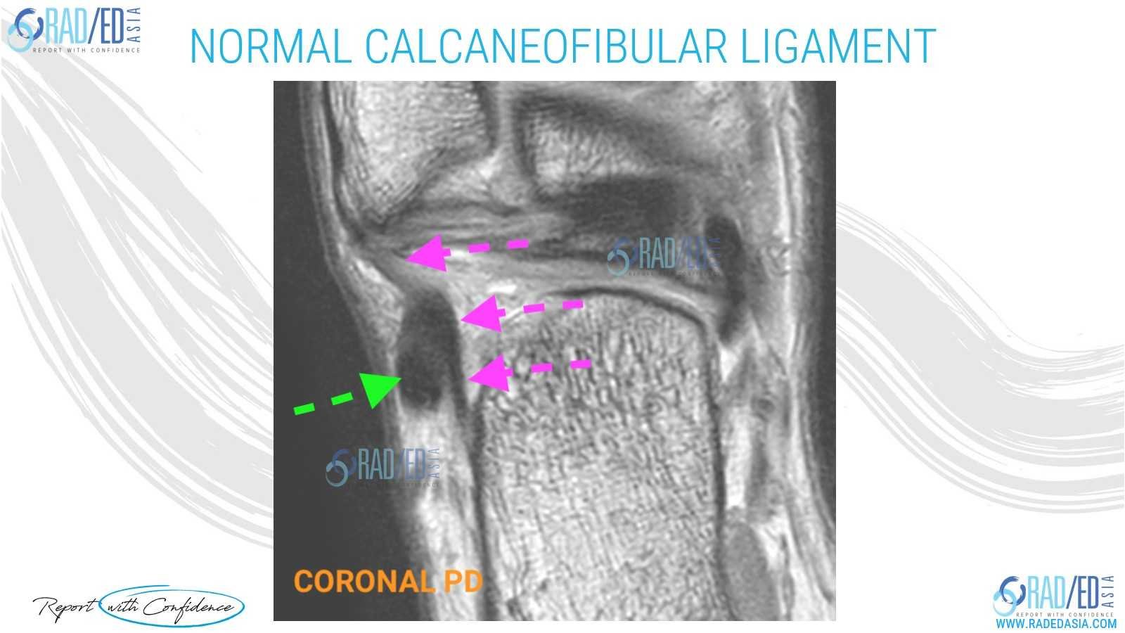
The normal MRI appearance of the Calcaneofibular ligament is a thin low signal structure that extends from the calcaneum to the fibula and passes deep to the peroneal tendons.![]()
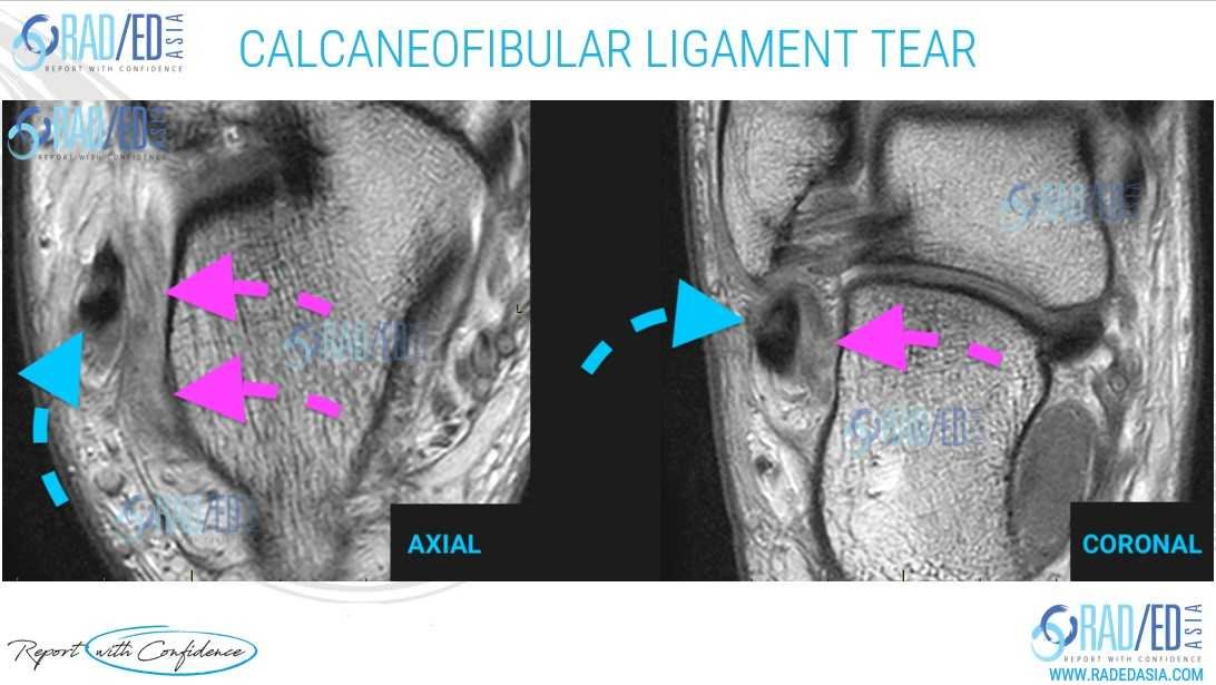
An acute high-grade tear of the Calcaneofibular ligament appears as thickening, hyper-intensity, and ill definition of the ligament.
![]()
The Peroneal tendons on an MRI can serve as an anatomical landmark to locate the Calcaneofibular ligamentFrom International Journal of Physical Medicine & Rehabilitation.![]()
A normal Calcaneofibular ligament appears as a thin low signal structure on an MRI.
![]()

