Dr. Ravi Padmanabhan

proximal tibiofibular joint ligament anatomy mri

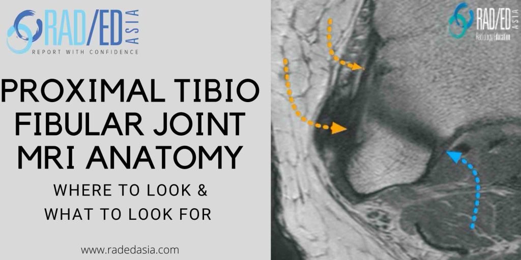
MRI KNEE ANATOMY: PROXIMAL TIBIOFIBULAR JOINT

PROXIMAL TIBIOFIBULAR JOINT MRI ANATOMY MRI PROXIMAL TIBIO FIBULAR JOINT ANATOMY The proximal tibiofibular joint is often overlooked when assessing a knee MRI. This is the first in a series on MRI of the proximal tibiofibular joint (PTFJ) beginning with the anatomy of the ligaments that stabilize the PTFJ. WHAT'S THE PROBLEM Often overlooked when …

MRI KNEE ANATOMY: PROXIMAL TIBIOFIBULAR JOINT Leer más »

spine facet joint mri online radiology courses

SPINE MRI NERVE ROOT COMPRESSION ONLINE RADIOLOGY COURSE

KEY POINTS FROM OUR SPINE MRI COURSES Spine MRI Nerve Root Compression Online Radiology Course Another three quick images with some basic key points from our online spine mri courses. Here we look at how to evaluate and describe a disc that extends into the foramen and its relationship to the exiting foraminal nerve looking …

SPINE MRI NERVE ROOT COMPRESSION ONLINE RADIOLOGY COURSE Leer más »

sesamoid osteonecrosis mri radiology

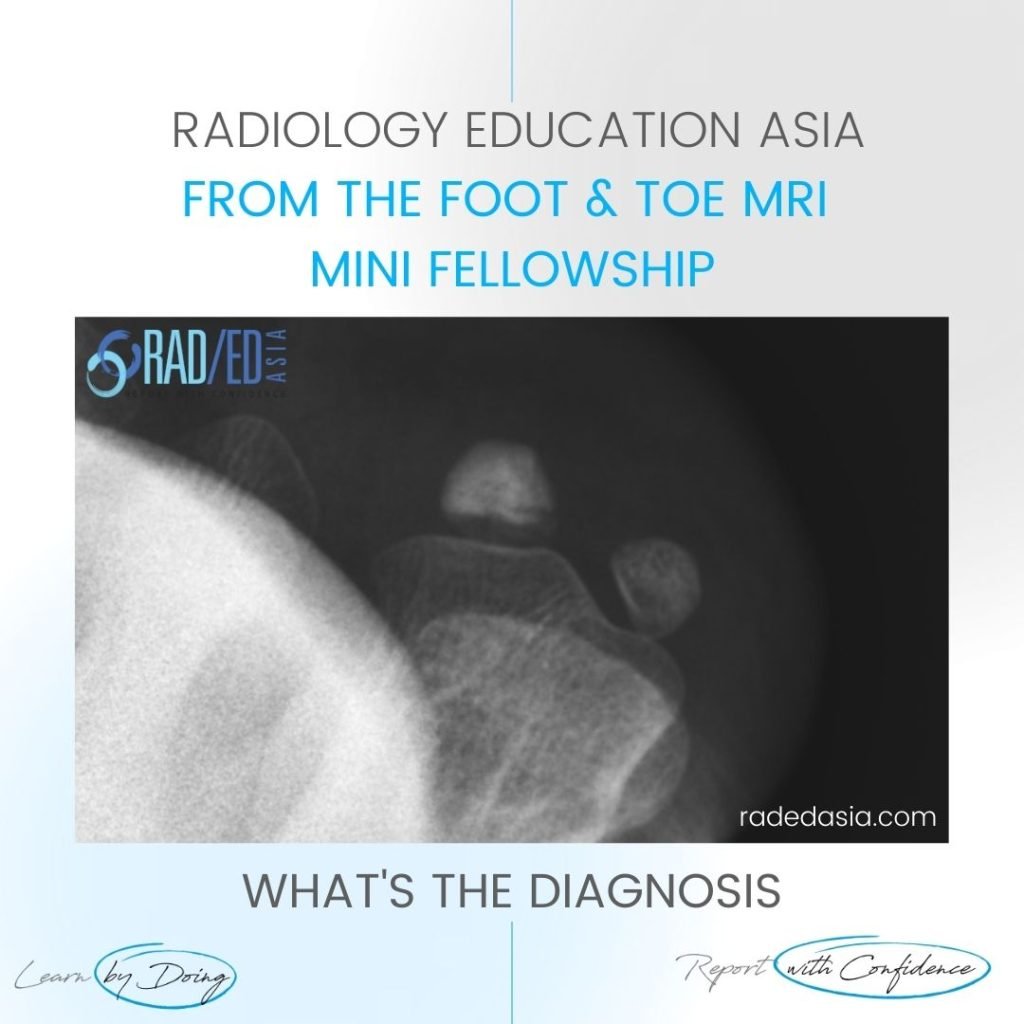
SESAMOID OSTEONECROSIS RADIOLOGY XRAY MRI

SESAMOID OSTEONECROSIS XRAY CT MRI: OVERVIEW OSTEONECROSIS OF THE SESAMOID Osteonecrosis of the sesamoids of the 1st metatarsal is not common but important that it’s recognized. As it progresses fracture, fragmentation and collapse of the sesamoid can occur. SESAMOID OSTEONECROSIS XRAY CT MRI: WHAT TO LOOK FOR SESAMOID OSTEONECROSIS XRAY: WHAT TO LOOK FOR If …

SESAMOID OSTEONECROSIS RADIOLOGY XRAY MRI Leer más »

mri-spine-how-to-mri-facet-cysts-spine-learn-spine-mri-online-radiology-education-asia

MRI LUMBAR FACET JOINT CYST RADIOLOGY: WHERE TO LOOK AND WHAT TO LOOK FOR (VIDEO)

LUMBAR SPINE FACET JOINT SYNOVIAL CYSTS MRI AND CT Lumbar Facet Joint Cysts MRI and CT: MRI Spine How to Assess for Facet Cysts and where to look. Today’s Tuesday Tip is a video on MRI of the Spine and Facet Cysts and in particular the MRI Spine Anatomy that relates to Spine Facet Cysts. …

MRI LUMBAR FACET JOINT CYST RADIOLOGY: WHERE TO LOOK AND WHAT TO LOOK FOR (VIDEO) Leer más »

learn-wrist-hand-elbow-mri-course-conference-abudhabi-dubai-uae-radedasia

WRIST, HAND AND ELBOW MRI CONFERENCE COURSE ABU DHABI UAE

WRIST, HAND & ELBOW MSK MRI CONFERENCE COURSE: Learn to Assess and Report with Confidence REGISTRATION CLOSED NOW Our new WRIST, HAND & ELBOW MRI MINI FELLOWSHIP is a Guided Hands-on Imaging Course to learn how to Assess and Report MRI Wrist, Hand & Elbow MRI More Confidently will be in Abu Dhabi, UAE on …

WRIST, HAND AND ELBOW MRI CONFERENCE COURSE ABU DHABI UAE Leer más »

mri periosteum callus periosteal new bone radedasia radiology education

PERIOSTEAL REACTION RADIOLOGY MRI: THE RANGE OF TRAUMATIC PERIOSTEAL CHANGES ON MRI

PERIOSTEAL REACTION RADIOLOGY MRI TRAUMATIC PERIOSTITIS & PERIOSTEAL CHANGE WHAT DOES PERIOSTEAL CHANGE SECONDARY TO TRAUMA LOOK LIKE ON MRI There are a number of appearances of periosteal reaction on MRI secondary to trauma that we can see and report. The Histology…Yes Histology!: It helps to have a simple understanding of the histology of the …

PERIOSTEAL REACTION RADIOLOGY MRI: THE RANGE OF TRAUMATIC PERIOSTEAL CHANGES ON MRI Leer más »

mri gleno humeral ligaments shoulder superior SGHL radiology education asia

GLENOHUMERAL LIGAMENTS MRI ANATOMY: SUPERIOR GLENO HUMERAL LIGAMENT SGHL

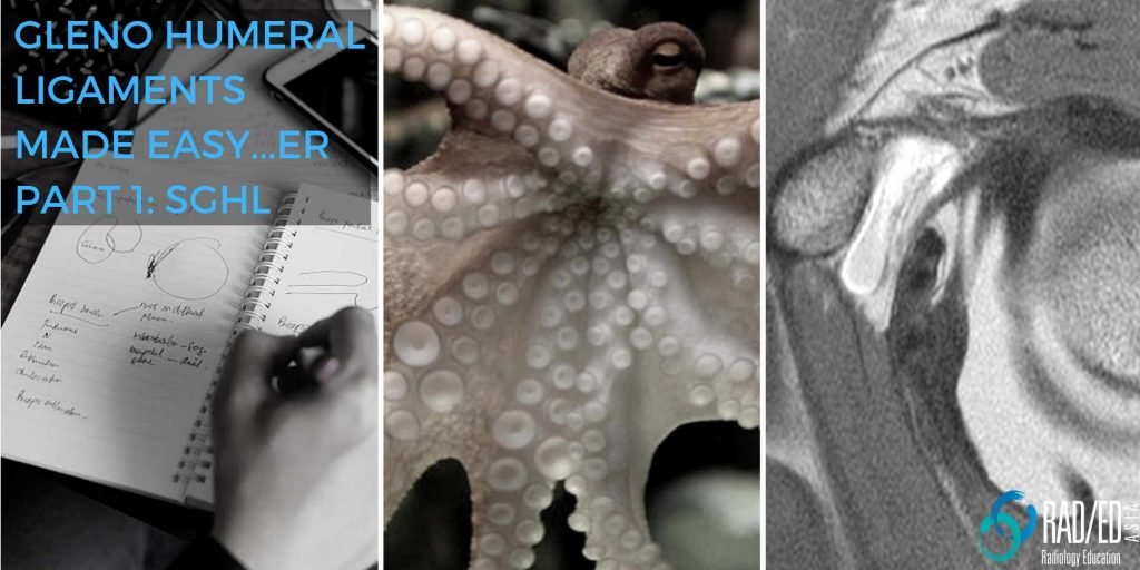
SUPERIOR GLENOHUMERAL LIGAMENT SGHL SGHL: How to identify the Gleno Humeral Ligaments made Easy…er Identifying the gleno humeral ligaments on MRI can be challenging as they are small and on lower field strength scanners there is inadequate resolution to see them properly. However, if we understand the anatomy of the ligaments, it becomes easier to …

GLENOHUMERAL LIGAMENTS MRI ANATOMY: SUPERIOR GLENO HUMERAL LIGAMENT SGHL Leer más »

mri-subscapularis-tendon-tear-atrophy-fatty-infiltration-biceps-subluxation-dislocation-radedasia

MRI OF SUBSCAPULARIS TEARS, ATROPHY, FATTY INFILTRATION, & BICEPS DISLOCATION

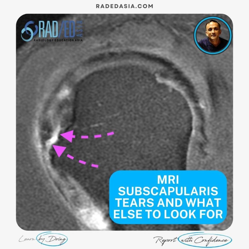
WHAT TO LOOK FOR ON MRI IN SUBSCAPULARIS TEARS, ATROPHY, FATTY INFILTRATION, & BICEPS DISLOCATION MRI Assessment of the subscapularis tendon is not just about looking for tears. This post looks at the four things that should be assessed.  The MRI appearance of partial and full-thickness tears.   Then assessing for muscle atrophy and fatty …

MRI OF SUBSCAPULARIS TEARS, ATROPHY, FATTY INFILTRATION, & BICEPS DISLOCATION Leer más »

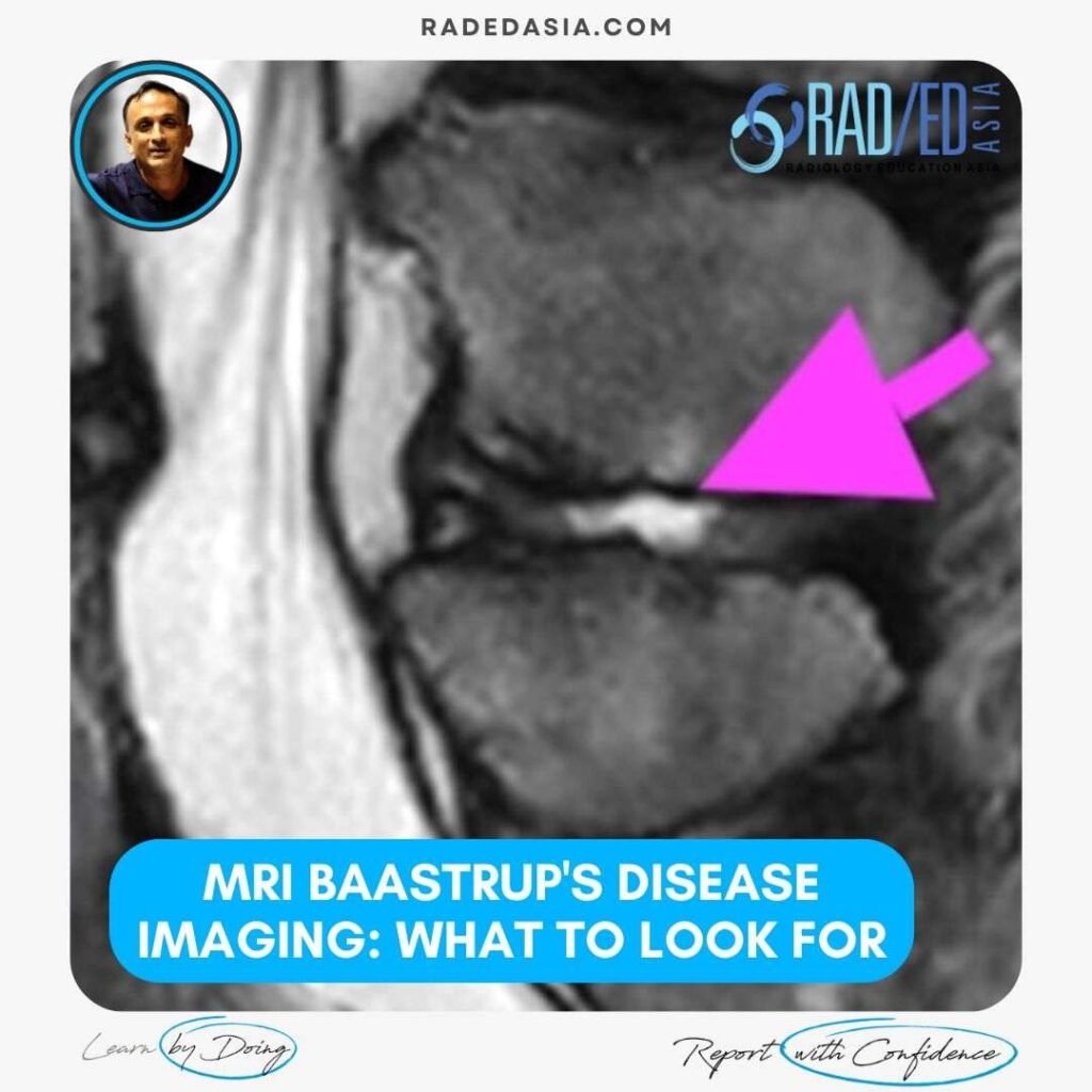
mri-baastrup's-disease-imaging-findings-what-to-look-for-radedasia

MRI BAASTRUP’S DISEASE: WHAT TO LOOK FOR

BAASTRUP'S DISEASE: WHAT TO LOOK FOR ON MRI Baastrup’s disease is caused by the friction of two spinous processes rubbing against each other and can result in central back pain but its difficult to diagnose without imaging. In the previous post we looked at the CT Findings in Baastrup’s Disease and this post looks at …

MRI BAASTRUP’S DISEASE: WHAT TO LOOK FOR Leer más »

Este sitio está destinado únicamente a profesionales médicos. El uso de este sitio se rige por nuestra Términos de servicio y Declaracion de privacidad que se puede encontrar haciendo clic en los enlaces. Acepte antes de continuar con el sitio web.
error: Clic derecho no disponible.