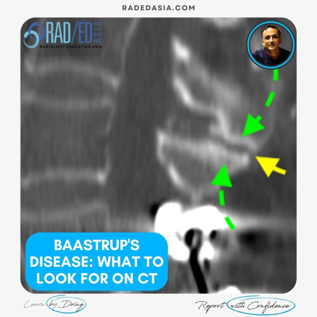
Post Blog

Blog Posts

mri knee meniscus degeneration maceration flap tear extrusion meniscocapsular separation radedasia

A FEW MRI MENISCUS ABNORMALITIES 2: MRI MENISCUS DEGENERATION MACERATION FLAP TEAR EXTRUSION AND MENISCOCAPSULAR SEPARATION

MENISCUS ABNORMALITIES 2 KNEE MRI MRI MENISCUS ABNORMALITIES A few more MRI Meniscus abnormalities such as meniscus maceration, flap tears, extrusion and meniscocapsular separation. MRI MENISCUS ABNORMALITIES MENISCO CAPSULAR SEPARATION MRI Meniscocapsular separation is a separation of the attachment of the external margin of the body of the medial meniscus from the Posterior Oblique ligament. …

A FEW MRI MENISCUS ABNORMALITIES 2: MRI MENISCUS DEGENERATION MACERATION FLAP TEAR EXTRUSION AND MENISCOCAPSULAR SEPARATION Leer más »

ankylosing spondylitis spine ct radiology

A FEW SPINAL CHANGES IN ANKYLOSING SPONDYLITIS

ANKYLOSING SPONDYLITIS SPINAL CHANGES CT & MRI Findings in Ankylosing Spondylitis of the spine Ankylosing spondylitis (AS), in the spine can involve the anterior and posterior elements. It usually commences anteriorly and as the disease progresses, posterior changes are seen. In these images we look at: Corner erosions and Sclerosis. Syndesmophyte formation between vertebrae. Fusion: …

A FEW SPINAL CHANGES IN ANKYLOSING SPONDYLITIS Leer más »

mri knee meniscus tears cysts pathologies images

A FEW MRI MENISCUS ABNORMALITIES: MENISCUS TEAR, INTRA MENISCAL CYST AND VARIANTS THAT LOOK LIKE TEARS

MENISCUS ABNORMALITIES KNEE MRI MRI MENISCUS ABNORMALITIES A few MRI Meniscus abnormalities such as Meniscus tear, Intra meniscal cyst and variants that look like tears. MRI MENISCUS ABNORMALITIES MRI MENISCUS INTRAMENISCAL CYST WHAT ARE THE FINDINGS? Cystic structure (Pink arrow) present within the anterior horn. WHAT’S THE Dx? INTRAMENISCAL CYST Extensive intra meniscal cyst anterior …

A FEW MRI MENISCUS ABNORMALITIES: MENISCUS TEAR, INTRA MENISCAL CYST AND VARIANTS THAT LOOK LIKE TEARS Leer más »

mri achilles tendon abnormalities ankle radiology

MRI OF A FEW ACHILLES TENDON ABNORMALITIES: INSERTIONAL TENDONOPATHY TENDONOSIS, BURSA & RUPTURE

ACHILLES TENDON ABNORMALITIES ANKLE MRI ACHILLES TENDON ABNORMALITIES A few MRI images of Achilles pathology from all the findings seen in Achilles Insertional Tendonopathy, Retrocalcaneal Bursitis, Retroachilles Bursitis to MRI of Achilles Rupture. MRI ACHILLES TENDON ABNORMALITIES MRI ACHILLES INSERTIONAL TENDONOPATHY: WHAT TO LOOK FOR MRI findings in Insertional Achilles Tendinopathy: A few findings to …

MRI OF A FEW ACHILLES TENDON ABNORMALITIES: INSERTIONAL TENDONOPATHY TENDONOSIS, BURSA & RUPTURE Leer más »

sesamoid osteonecrosis mri radiology

SESAMOID OSTEONECROSIS RADIOLOGY XRAY MRI

SESAMOID OSTEONECROSIS XRAY CT MRI: OVERVIEW OSTEONECROSIS OF THE SESAMOID Osteonecrosis of the sesamoids of the 1st metatarsal is not common but important that it’s recognized. As it progresses fracture, fragmentation and collapse of the sesamoid can occur. SESAMOID OSTEONECROSIS XRAY CT MRI: WHAT TO LOOK FOR SESAMOID OSTEONECROSIS XRAY: WHAT TO LOOK FOR If …

SESAMOID OSTEONECROSIS RADIOLOGY XRAY MRI Leer más »

pyriformis syndrome pudendal nerve quadratus femoris ischiofemoral impingement ultrasound scan inject radedasia

ULTRASOUND PAIN MANAGEMENT HOW TO SCAN AND INJECT FOR PIRIFORMIS SYNDROME, PUDENDAL NERVE, QUADRATUS FEMORIS & ISCHIOFEMORAL IMPINGEMENT

STEP BY STEP GUIDE ON HOW TO SCAN AND INJECT THE GLUTEAL REGION FOR PAIN MANAGEMENT HOW TO SCAN AND INJECT THE GLUTEAL REGION FOR PAIN MANAGEMENT   In this video by Dr Rajendra Sahoo Pain Management Specialist at KIMS Hospital and Neuron Pain Clinic we look at the role of ultrasound in the diagnosis …

ULTRASOUND PAIN MANAGEMENT HOW TO SCAN AND INJECT FOR PIRIFORMIS SYNDROME, PUDENDAL NERVE, QUADRATUS FEMORIS & ISCHIOFEMORAL IMPINGEMENT Leer más »

meniscal ossicles mri knee radiology education asia

MENISCAL OSSICLES: POSTERIOR HORN MENISCUS PART 3

MENISCAL OSSICLES: POSTERIOR HORN MENISCUS MENISCAL OSSICLES: WHAT ARE THEY Meniscal ossicles are areas of ossification that occur within the meniscus. WHY DO THEY OCCCUR Two possible causes are given: They are a Result of Post traumatic metaplasia and heterotopic ossification of a meniscal tear (either posterior horn or root). This is the most likely …

MENISCAL OSSICLES: POSTERIOR HORN MENISCUS PART 3 Leer más »

acromial spur mri shoulder radiology education asia

ACROMION / ACROMIAL SPURS ON MRI: WHERE TO LOOK AND WHAT TO LOOK FOR

ACROMION SPURS ON MRI: 3 APPEARANCES ON MRI Acromial / Acromion spurs can be quite small and difficult to visualize on MRI. They are enthesophytes that arise from traction at the attachment sites of the Coraco Acromial Ligament and Deltoid. This post looks at the three locations and most common appearances of spurs arising from …

ACROMION / ACROMIAL SPURS ON MRI: WHERE TO LOOK AND WHAT TO LOOK FOR Leer más »

Este sitio está destinado únicamente a profesionales médicos. El uso de este sitio se rige por nuestra Términos de servicio y Declaracion de privacidad que se puede encontrar haciendo clic en los enlaces. Acepte antes de continuar con el sitio web.
error: Clic derecho no disponible.