Post Blog

Blog Posts

iliotibial band mri anatomy syndrome trauma mri knee radedasia

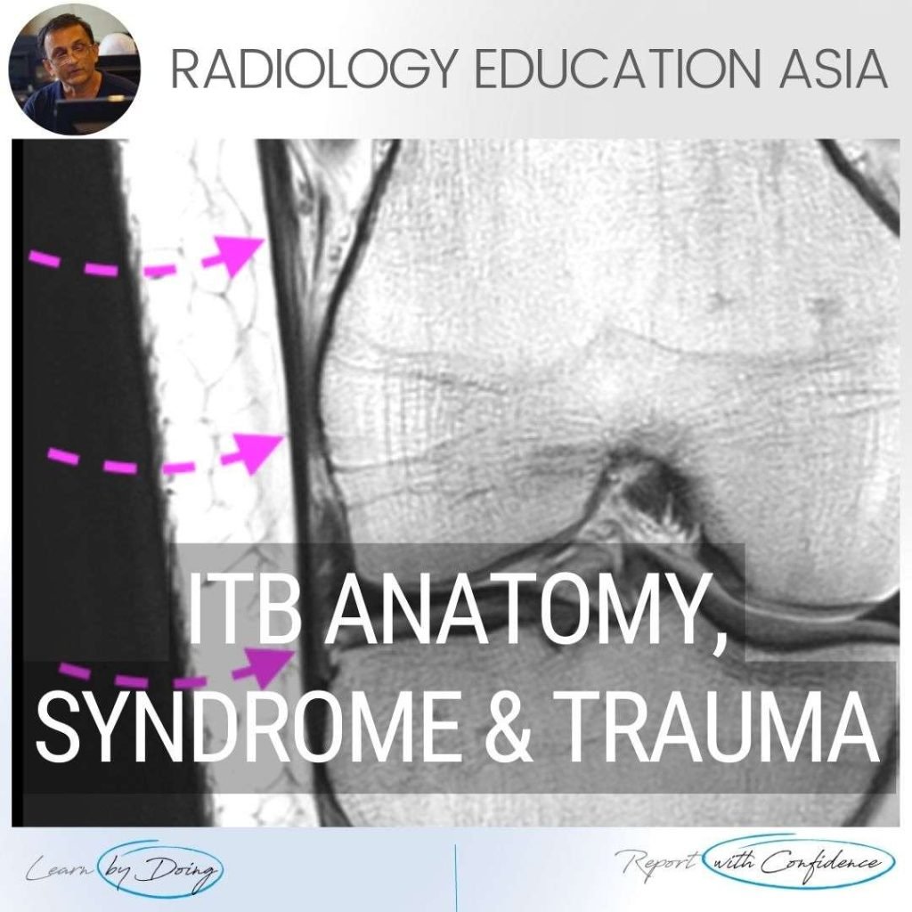
ILIOTIBIAL BAND MRI ANATOMY, SYNDROME, TRAUMA KNEE RADIOLOGY

ILIOTIBIAL BAND MRI ANATOMY, SYNDROME, TRAUMA Finding and assessing the Iliotibial Band (ITB) on MRI of the Knee is the first structure we assess when looking at the lateral side of the Knee.  In these three posts we look at: The normal MRI anatomy of the ITB. MRI of Iliotibial Band Syndrome (ITB Friction Syndrome) …

ILIOTIBIAL BAND MRI ANATOMY, SYNDROME, TRAUMA KNEE RADIOLOGY Leer más »

shoulder labrum mri variant anatomy bicipito labrum complex radedasia

SHOULDER LABRUM MRI VARIANT ANATOMY BICIPITO LABRUM COMPLEX: Don’t call these three appearances of the labrum a tear.

SHOULDER MRI LABRUM MRI VARIANT ANATOMY There are a number of normal appearances of the Bicipito Labral Complex which can mistakenly be called tears.   Here is what they are and where to look for them.   (These posts are Part of our Quick Pick Series with concise information from the Journals I read). CLICK …

SHOULDER LABRUM MRI VARIANT ANATOMY BICIPITO LABRUM COMPLEX: Don’t call these three appearances of the labrum a tear. Leer más »

mri musculotendinous junction cysts and delaminating rotator cuff tears

MRI DELAMINATION ROTATOR CUFF TEARS MUSCULOTENDINOUS JUNCTION CYSTS

MRI DELAMINATING ROTATOR CUFF TEARS & MUSCULOTENDINOUS JUNCTION CYSTS On MRI Rotator cuff delaminating tears can result in Musculotendinous Junction Cysts.   These Musculotendinous junction cysts are seen in the rotator cuff tendons, mostly in Supraspinatus and less commonly in infraspinatus.   In this post we look at the appearance of both Delaminating Rotator cuff …

MRI DELAMINATION ROTATOR CUFF TEARS MUSCULOTENDINOUS JUNCTION CYSTS Leer más »

spine mri msk online radiology courses radedasia

LUMBAR SPINE ENHANCEMENT MRI ONLINE RADIOLOGY COURSES: KEY POINTS FROM OUR SPINE MRI COURSES

KEY POINTS FROM OUR SPINE MRI COURSES Spine MRI Online Radiology Course: Normal Spinal Enhancement Another three quick images with some basic key points from our online spine MRI courses. Here we look at areas of normal enhancement that can be mistaken for pathology: Normal enhancement of the anterior epidural plexus which can be mistaken …

LUMBAR SPINE ENHANCEMENT MRI ONLINE RADIOLOGY COURSES: KEY POINTS FROM OUR SPINE MRI COURSES Leer más »

sacral fracture lumbar mri radiology education asia

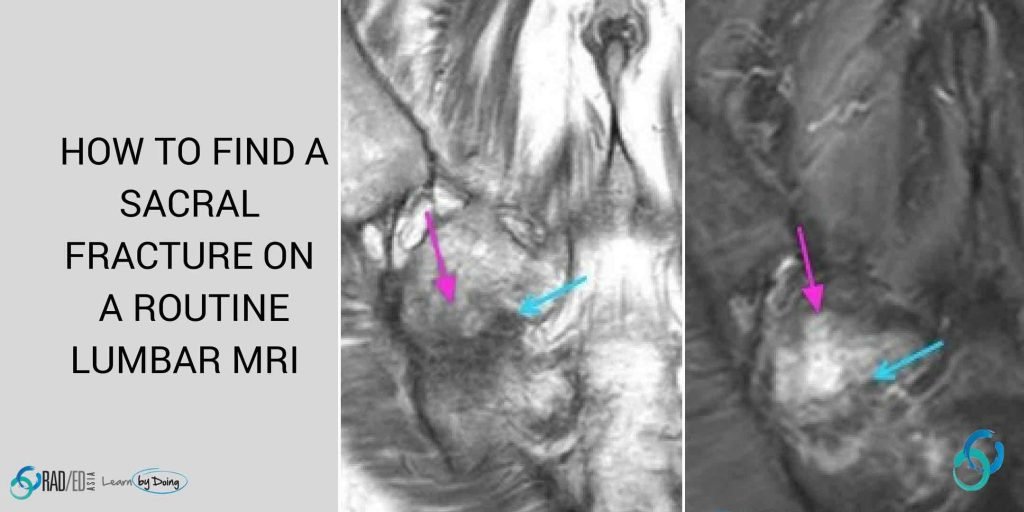
HOW TO FIND A SACRAL FRACTURE ON A LUMBAR MRI

SACRAL FRACTURE MRI: HOW TO FIND ON A LUMBAR MRI HOW TO FIND SACRAL FRACTURE ON LUMBAR MRI When the clinical examination and referral notes are inadequate (Back Pain F.I.) the cause of the pain may well be sacral rather than lumbar. On a standard lumbar spine MRI with axial and sagittal scans,  the sacrum …

HOW TO FIND A SACRAL FRACTURE ON A LUMBAR MRI Leer más »

synovitis mri appearances

SYNOVITIS MRI FINDINGS KNEE WHAT DOES IT LOOK LIKE

SYNOVITIS MRI FINDINGS SYNOVITIS MRI FINDINGS: WHAT DOES IT LOOK LIKE   Synovitis on MRI, like the many faced god in Game of Thrones, can have many appearances. However, like we saw in the article Tendinosis: Same Same But Different once you recognizes the different appearances, it’s the same pattern for each joint. Synovitis is …

SYNOVITIS MRI FINDINGS KNEE WHAT DOES IT LOOK LIKE Leer más »

plantar fascia plantar fasica tears plantar fasciitis plantar fascia anatomy bands mri plantar fasci

MRI PLANTAR FASCIA TEAR PLANTAR FASCIITIS MRI (VIDEO)

MRI PLANTAR FASCIA TEAR & PLANTAR FASCIITIS: WHAT TO LOOK FOR MRI PLANTAR FASCIA TEAR PLANTAR  FASCIITIS MRI   In this video we are looking at the MRI findings in Plantar Fasciitis and Plantar Fascia Tears. Previously we have looked at MRI of Plantar Fascia Anatomy and Where an What to assess in the Plantar …

MRI PLANTAR FASCIA TEAR PLANTAR FASCIITIS MRI (VIDEO) Leer más »

mri spine rheumatoid arthritis basilar invagination radiology education asia

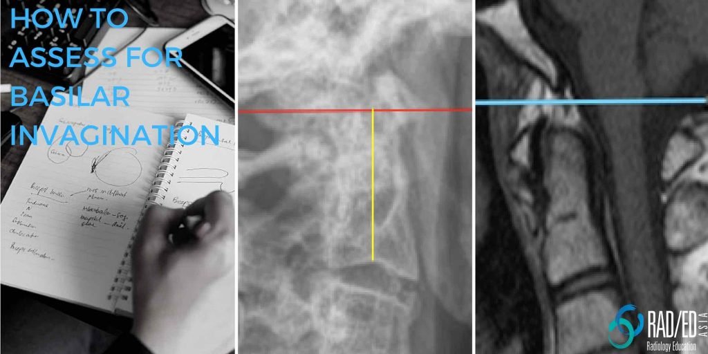
BASILAR INVAGINATION RADIOLOGY RHEUMATOID ARTHRITIS FIVE WAYS TO ASSESS

BASILAR INVAGINATION MRI RHEUMATOID ARTHRITIS WHAT IS BASILAR INVAGINATION / IMPRESSION BASILAR INVAGINATION RADIOLOGY RHEUMATOID ARTHRITIS: Subluxation at C1/2 in Rheumatoid Arthritis can occur in three directions. Axial (Horizontal). Vertical and, Lateral. Vertical subluxation is when C2 moves proximally towards the foramen magnum and is called Basilar Invagination or Basilar Impression. Basilar Invagination is when …

BASILAR INVAGINATION RADIOLOGY RHEUMATOID ARTHRITIS FIVE WAYS TO ASSESS Leer más »

synovitis mri ighl shoulder

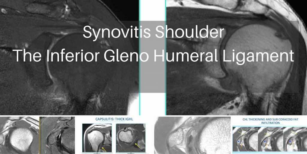
MRI SYNOVITIS SPECIFIC SITES IN THE SHOULDER: THE INFERIOR GLENOHUMERAL LIGAMENT IGHL

MRI Shoulder Synovitis Capsulitis: Inferior Glenohumeral Ligament IGHL Capsulo Synovitis MRI Capsulo Synovitis IGHL. At our MRI workshops we have an Open Mic policy. People are encouraged to interrupt and ask questions at anytime because its best to clear doubts at the time topics are being discussed rather than wait till the end or never …

MRI SYNOVITIS SPECIFIC SITES IN THE SHOULDER: THE INFERIOR GLENOHUMERAL LIGAMENT IGHL Leer más »

Este sitio está destinado únicamente a profesionales médicos. El uso de este sitio se rige por nuestra Términos de servicio y Declaracion de privacidad que se puede encontrar haciendo clic en los enlaces. Acepte antes de continuar con el sitio web.
error: Clic derecho no disponible.