
COLUMNA VERTEBRAL
Crowned dens is a manifestation of Calcium Pyrophosphate deposition diseases (CPPD) in the cervical spine.
![]()
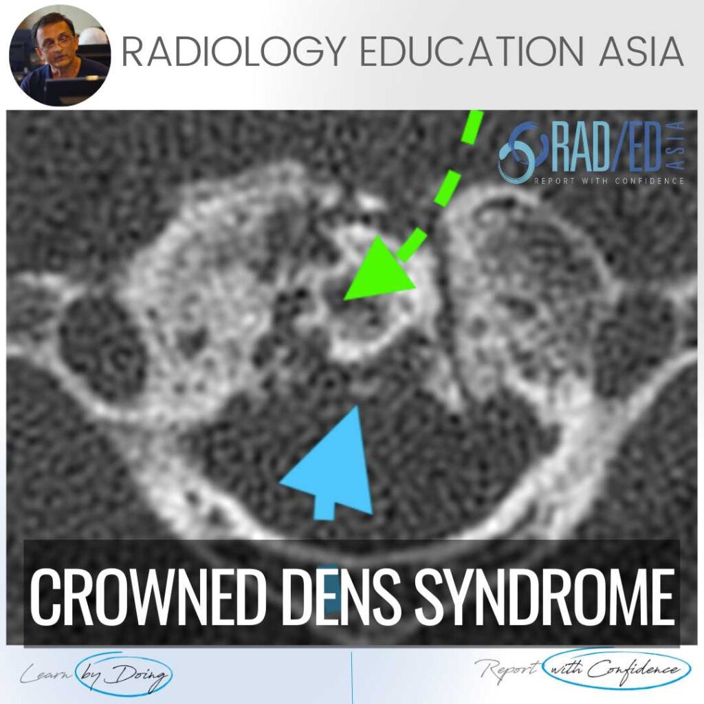
Peri odontoid deposition of Calcium Pyrophosphate crystals.
The diagnosis is easier on CT as high density of calcium is seen around the dens.

Image above: Increased density ( Pink arrows) centred in the region of the transverse ligament.
 Image above: Axial T2 scans demonstrates low signal thickening of soft tissue (Pink arrow) centered in the region of the transverse ligament. Green arrow Dens.
Sagittal Gradient echo sequence demonstrates areas of low signal (Yellow arrow) in keeping with calcification.
Image above: Axial T2 scans demonstrates low signal thickening of soft tissue (Pink arrow) centered in the region of the transverse ligament. Green arrow Dens.
Sagittal Gradient echo sequence demonstrates areas of low signal (Yellow arrow) in keeping with calcification.
 Thank you to Dr Joe Thomas Chief Rheumatologist, Aster Medcity Kochi, for his input in the case. Rheumatology Imaging is made much easier with the guidance of a Rheumatologist in the clinical aspects and what they need from a report.
We have Dr Thomas joining us in presenting the Spine Arthropathy & Spondyloarthropathy course to give us the important clinical aspects we need to know as radiologists and also what a Rheumatologist wants from our reports. Get More information by clicking on the images below.
Thank you to Dr Joe Thomas Chief Rheumatologist, Aster Medcity Kochi, for his input in the case. Rheumatology Imaging is made much easier with the guidance of a Rheumatologist in the clinical aspects and what they need from a report.
We have Dr Thomas joining us in presenting the Spine Arthropathy & Spondyloarthropathy course to give us the important clinical aspects we need to know as radiologists and also what a Rheumatologist wants from our reports. Get More information by clicking on the images below. 


