ISCHIOFEMORAL IMPINGEMENT MRI
Ischiofemoral Impingement is sometimes missed in Hip MRI because our focus is too narrowly centered on the joint or hamstrings. When assessing the hip joint it helps to organize the evaluation into compartments like the joint, trochanteric region, hamstrings and surrounding muscles and tendons so that each area is specifically assessed.
Like meniscal root tears, ischiofemoral impingement is one of those things that you need to look for specifically rather than as part of a general assessment of the hip as we scroll through the scan.
Ischiofemoral impingement can result in deep posterior buttock pain and due to is the compression of the Quadratus Femoris Muscle between the Ischial Tuberosity and the Femur, at the level of the lesser trochanter. This is due to the narrowing of the space where the Quadratus Femoris lies with compression of the muscle between the ischial tuberosity and the lesser trochanter.
Narrowing and Compression can result in
- Quadratus Femoris Oedema.
- Quadratus femoris Tears.
- Quadratus Femoris Atrophy.
We need to look for
1. Changes to the Quadratus Femoris
2. Narrowing of the space where Quadratus Femoris is located.
The Quadratus Femoris lies in the space between the ischial tuberosity and the lesser trochanter.
Image Above: Ischiofemoral Impingement MRI Anatomy. Ischial Tuberosity (Pink Arrow), Lesser Tuberosity (Orange Arrow), Iliopsoas Tendon (Blue arrow), Hamstrings (Yellow Arrow), Quadratus Femoris (Green Dots)
Two areas can be measured for narrowing of the space where the Quadratus Femoris lies, although its often more practical to " eyeball" the region and decide if it looks narrowed or not.
Additionally a normal size Ischiofemoral space can be seen with Quadratus Femoris changes of impingement as we are scanning the patient at rest and narrowing and compression may only occur when the patient is in another position.
Ischiofemoral space: Measure the smallest distance between the Medial margin of the lesser trochanter and the lateral margin of ischial tuberosity
Normal Controls 23 +/-8mm
IschioFemoral Impingement: 13 +/-5 mm
Cutoff: < 17mm
Quadratus femoris space : Measure the smallest distance between the Lesser Trochanter and the Hamstrings.
Controls: 12 +/-3mm
IschioFemoral Impingement:7 +/-4mm
Cutoff: <8mm
(All measurements from AJR: 193 July 2009 but small sample size).
Image Above: Ischiofemoral Impingement MRI where to measure. IschioFemoral Space from Ischial Tuberosity to Lesser Trochanter (Blue Line) Quadratus Femoris Space Shortest distance from Anterolateral Margin Hamstrings to Lesser Trochanter (Pink Line).
Image Above: Ischiofemoral Impingement MRI The only finding suggesting impingement may be only Quadratus femoris oedema increased PDFS signal (Blue arrow) without a tear.
Ischial Tuberosity (Pink Arrow), Lesser Tuberosity (Orange Arrow), Hamstrings (Yellow Arrow).
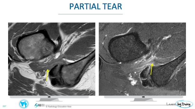
Image Above: Ischiofemoral Impingement MRI Quadratus femoris is partially torn.
Image Above: Ischiofemoral Impingement MRI Quadratus femoris is thinned and reduced volume in keeping with partial tearing and atrophy (Blue arrow), Ischial Tuberosity (Pink Arrow), Lesser Tuberosity (Orange Arrow), Hamstrings (Yellow Arrow).
Learn more about this condition & How best to report it in more detail in
our Online Guided HIP MRI Mini Fellowship.
More by clicking on the images below.
For all our other current MSK MRI & Spine MRI
Online Guided Mini Fellowships.
Click on the image below for more information.
- Join our WhatsApp RadEdAsia community for regular educational posts at this link: https://bit.ly/radedasiacommunity
- Get our weekly email with all our educational posts: https://bit.ly/whathappendthisweek
#radedasia #mri #mskmri #radiología